

Sabato 27 ottobre 2012 - Pacini Editore
Sabato 27 ottobre 2012 - Pacini Editore
Sabato 27 ottobre 2012 - Pacini Editore

Create successful ePaper yourself
Turn your PDF publications into a flip-book with our unique Google optimized e-Paper software.
288<br />
Methods. In this study we examined a series of 192 colorectal<br />
carcinomas surgically resected from January 2003 to<br />
June 2010. Histological evaluation was done using standard<br />
histologic sections stained with hematoxylin and eosin. The<br />
tumors were widely sampled, with a number of tissue blocks<br />
of approximately one per cm in largest tumor diameter. A<br />
carcinoma was classified as micropapillary when micropapillary<br />
tufts lacking true fibrovascular cores and surrounded by<br />
empty lacunar spaces represented at least 5% of the tumor area<br />
in histologic sections.<br />
Results. 29 of the 192 tumors (15.1%) were classified as MPC.<br />
In most cases the MP component represented < 30% of the<br />
tumor volume. MP carcinomas were observed more often in<br />
males than females (19.8% vs. 8.6%, P = 0.04). In contrast patient’s<br />
age was not significantly related with the presence of MP<br />
features. A highly significant relationship was evident between<br />
MP component and tumor site. A MP component was present<br />
in <strong>27</strong>.9% of tumors located in the rectum, in 20% of tumors of<br />
the left colon (descending and sigmoid colon) and in 7.1% of<br />
tumors arising in the proximal colon (right and transverse colon)<br />
(P = 0.003). MPCs were more often in advanced stages (III<br />
and IV) compared to conventional adenocarcinomas (72.4% vs.<br />
34.4%, P < 0.001). In addition, MPCs showed a more advanced<br />
pT category (P = 0.03), more frequent involvement of the serosa<br />
(41.4% vs. 21.5%, P = 0.03) and a much greater propensity<br />
to lymph node metastasis (69.0% vs. 31.9%, P < 0.001) than<br />
conventional adenocarcinomas. 48.3% of MPCs showed 4 or<br />
more lymph node metastases, while only 15.3% of common<br />
adenocarcinomas were classified as pN2. The frequency of distant<br />
metastases at diagnosis was higher in the MPCs (17.2% vs.<br />
9.8%), but the difference did not reach statistical significance.<br />
Tumors with a MP component also showed more frequently<br />
an infiltrative pattern of growth (47.4% vs. 13.8%, P = 0.002)<br />
and extramural venous invasion (37.9% vs. 16.6%, P = 0.01).<br />
Finally in our study 24 of the 29 MPCs (82.8%) were classified<br />
as poorly differentiated (high grade), while only 22.1% of conventional<br />
carcinomas were high grade (P < 0.001).<br />
Conclusions. The results we obtained confirm the characteristics<br />
of pathological aggressiveness of the MPCs reported in<br />
literature and highlight the high propensity to lymph node metastasis<br />
of these tumors. The prognostic significance of the MP<br />
histotype has been evaluated only in a limited number of studies.<br />
Given the association between MP component and lymph<br />
node metastasis, distant metastasis and other pathological<br />
parameters of aggressiveness, it is expected that this histological<br />
type is characterized by a generally poor prognosis, with<br />
survival rates lower than those observed in common adenocarcinomas<br />
and mucinous adenocarcinomas. Further studies<br />
are certainly necessary to verify if the MP histology represents<br />
a prognostic factor more important and reproducible than the<br />
degree of differentiation and to define the biomolecular and<br />
genetic basis of this tumour type.<br />
references<br />
1 Verdù M, et al. Clinicopathological and molecular characterization of<br />
colorectal micropapillary carcinoma. Mod Pathol 2011;24:729-38.<br />
2 Xu F, et al. Micropapillary component in colorectal carcinoma is<br />
associated with lymph node metastasis in T1 and T2 stages and<br />
decreased survival time in TNM stages I and II. Am J Surg Pathol<br />
2009;33:1287-92.<br />
3 Kim M-J, et al. Invasive colorectal micropapillary carcinoma: an<br />
aggressive variant of adenocarcinoma. Hum Pathol 2006;37:809-15.<br />
4 Haupt B, et al. Colorectal adenocarcinoma with micropapillary<br />
pattern and its association with lymph node metastasis. Mod Pathol<br />
2007;20:729-33.<br />
Sakamoto K. Primary invasive micropapilklary carcinoma of the colon.<br />
Histopathology 2005;47:479-84.<br />
CONGRESSO aNNualE di aNatOmia patOlOGiCa SiapEC – iap • fiRENzE, 25-<strong>27</strong> OttOBRE <strong>2012</strong><br />
Usefulness of anti-TCrGD antibody in gluten<br />
sensitivity diagnosis on formalin-fixed paraffinembedded<br />
biopsies<br />
S. Lonardi1 , V. Villanacci1 , A. Lanzini2 , F. Lanzarotto2 , L. Lorenzi1<br />
, U. Volta3 , F. Facchetti1 1 2 Department of Pathology, Gastroenterology Unit, Spedali Civili-University<br />
of Brescia, 3 Department of Clinical Medicine, St Orsola-Malpighi<br />
Hospital-University of Bologna, Bologna, Italy<br />
Introduction. Small bowel intraepithelial lymphocytosis (IL) may<br />
depend from different causes, including Gluten Sensitive Enteropathy<br />
(GSE). 1 Demonstration of increased number of duodenal<br />
T-cell receptor gamma-delta (TCRGD) positive intraepithelial<br />
lymphocytes (IELs) has been used to support GSE diagnosis on<br />
frozen material. 2-3 We have evaluated an anti-TCRGD antibody on<br />
formalin-fixed paraffin embedded (FFPE) small bowel biopsies.<br />
Material and methods. Anti-CD3 (clone SP7, 1:100) and anti-<br />
TCRGD (TCR CgM1, clone g3.20, 1:400) from Thermo Scientific,<br />
Fremont, CA) were applied by immunohistochemistry on 59 FFPE<br />
biopsies from 18 cases of celiac disease (CD) with mild/severe atrophy,<br />
19 cases of IL in CD patients on Gluten Free Diet (IL-GFD),<br />
14 cases of IL (6/14 with abnormal serology for GSE), and 8 controls<br />
(CTR) with mild duodenitis and negative CD serology and genotyping.<br />
Sections were digitalized with Aperio Scanscope (Nikon)<br />
and IELs/100 epithelial cells (EC) were counted in six high power<br />
fields. For statistic analysis the Mann-Whitney test was applied.<br />
Results. CD3+ IELs were significantly higher in CD (mean±SD<br />
52.0±15.3) (Figure 1A), IL-GFD (43.6±18.3) and IL (46.3±16.6)<br />
(Figure 2A) compared to CTR (24.1±7.6) (p = 0.0004, p = 0.0019<br />
and p = 0.0006, respectively) (Figure 3A)<br />
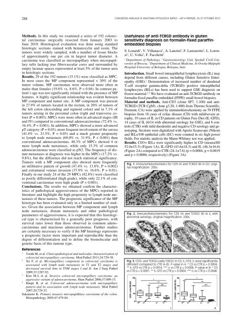
Fig. 1. immunohistochemistry for Cd3 (a) and tCRGd (B) in Cd; original<br />
magnification: 200x.<br />
Fig. 2. immunohistochemistry for Cd3 (a) and tCRGd (B) in il with<br />
abnormal serology; original magnification: 200x.<br />
Fig. 3. Cd3+ and tCRGd+cells/100 EC in Cd, il-Gfd, il were significantly<br />
different compared to CtR (a-B). p-value in a: * Cd vs CtR p = 0.0004,<br />
** il-Gfd vs CtR p = 0.0019, *** il vs CtR p = 0.0006. p-value in B: * Cd<br />
vs CtR p < 0.0001, ** il-Gfd vs CtR p = 0.0004, *** il vs CtR p = 0.0046.


