

Growth, Differentiation and Sexuality
Growth, Differentiation and Sexuality
Growth, Differentiation and Sexuality

You also want an ePaper? Increase the reach of your titles
YUMPU automatically turns print PDFs into web optimized ePapers that Google loves.
in membrane enlargement <strong>and</strong> exocytosis would<br />
occur.<br />
The apical vesicles are thought to arise in the<br />
endoplasmic reticulum far behind the tip, pass<br />
throughGolgiequivalents,<strong>and</strong>bethentransported<br />
vectorially to the apex where they fuse with the<br />
plasma membrane (see The Mycota, Vol. I, 1st edn.,<br />
Chap. 3). In their membranes, these vesicles may<br />
carry proteins destined for the plasma membrane,<br />
such as polysaccharide synthases, ion channels <strong>and</strong><br />
ATP-ase. In their lumina, they may carry some wall<br />
components, wall-modifying enzymes, as well as<br />
lytic enzymes destined for excretion to digest polymeric<br />
substrates. The cytological <strong>and</strong> enzymological<br />
evidence attesting to this view has been reviewed<br />
by Gooday <strong>and</strong> Gow (1990). As mentioned<br />
above, evidence is accumulating that chitin synthesis<br />
in yeast <strong>and</strong> at the hyphal tip is regulated by exo<strong>and</strong><br />
endocytosis of chitin synthase-containing particles<br />
(see The Mycota, Vol. III, 2nd edn., Chap. 14).<br />
B. TheHyphalWallasaBarrierforPassage<br />
of Proteins<br />
Thecellwallisthelastbarrierproteinssecretedinto<br />
the medium have to take. Slime mutants of N. crassa<br />
whicharedefectiveinwallsynthesisover-excrete<br />
enzymes, including invertase (Bigger et al. 1972;<br />
Casanova et al. 1987; Pietro et al. 1989), alkaline<br />
phosphatase (Burton <strong>and</strong> Metzenberg 1974), alkaline<br />
protease <strong>and</strong> aryl-β-glucosidase (Pietro et al.<br />
1989). This suggests that the wall acts as a barrier<br />
in protein excretion. Moreover, the problem of<br />
passage through the wall is compounded by the fact<br />
that excreted proteins are sometimes much larger<br />
than the estimated average pore size of the walls of<br />
yeasts (Scherrer et al. 1974; Cope 1980) <strong>and</strong> mycelial<br />
fungi (Trevithnick <strong>and</strong> Metzenberg 1966). These<br />
pore sizes were measured with isolated cell wall<br />
fractions, or determined by the solute exclusion<br />
method, using living hyphae (Money 1990).<br />
On the basis of the existence of a correlation<br />
between the sizes of excreted proteins <strong>and</strong> numbers<br />
of hyphal tips present, Chang <strong>and</strong> Trevithick<br />
(1974) have proposed that the proteins are excreted<br />
at the hyphal apices <strong>and</strong> that the nascent wall at the<br />
apex contains larger pores than the mature subapical<br />
wall. Because of the small proportion of apical<br />
wall material in wall preparations used for poresize<br />
measurements, such large pores would go undetected.<br />
The description of wall deposition at the<br />
extreme apex of the hypha, given in Sect. V., indeed<br />
Apical Wall Biogenesis 65<br />
indicates that the nature of the wall in that region<br />
differs completely from that in subapical regions; it<br />
can easily be envisaged that the assumed gel-like,<br />
highly hydrated condition of the wall at the extreme<br />
apex facilitates the diffusion of large proteins over<br />
this barrier. However, the measurements of Money<br />
(1990) on solute exclusion in living hyphae do not<br />
support this contention.<br />
An alternative is offered by the bulk-flow hypothesis<br />
proposed by Wessels (1990, 1993, 1999),<br />
which assumes that proteins secreted at the very<br />
tip are pushed through the wall from the inside to<br />
the outside by the accretion of plastic wall polymers<br />
during wall growth. A theoretical consideration on<br />
how a wall volume travels through an exp<strong>and</strong>ing<br />
wall at the apex is given by Green (1974). Considering<br />
the model for apical wall growth discussed<br />
in Sect. V., it is plausible that proteins extruded<br />
into the wall will be carried by the flow of plastic<br />
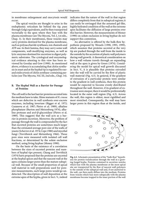
wall material (Fig. 4.3). In general, if the gradient<br />
of extrusion of a particular protein were similar<br />
to the gradient in wall synthesis, then this protein<br />
would be expected to become evenly distributed<br />
throughout the wall. However, if its gradient of extrusion<br />
were steeper, then it would be preferentially<br />
located in the outer wall region (Fig. 4.3). Across<br />
the wall, this region is oldest, most rigidified <strong>and</strong><br />
most stretched. Consequently, the wall may have<br />
larger pores in this region than at the inside, <strong>and</strong><br />
Fig. 4.3. Schematic presentation of the “bulk-flow” hypothesis<br />
for protein translocation through the wall in a growing<br />
fungal hypha. Proteins contained in secretion vesicles<br />
which fuse with the plasma membrane at the most apical<br />
site are transported by the flow of nascent wall polymers<br />
to the outside of the subapical wall <strong>and</strong>, if not anchored to<br />
the wall, can then easily diffuse into the medium. Proteins<br />
fromvesicleswhichfusemoresubapicallywiththeplasma<br />
membrane become trapped in the inner portion of the wall


