2011 (SBTE) 25th Annual Meeting Proceedings - International ...
2011 (SBTE) 25th Annual Meeting Proceedings - International ...
2011 (SBTE) 25th Annual Meeting Proceedings - International ...
Create successful ePaper yourself
Turn your PDF publications into a flip-book with our unique Google optimized e-Paper software.
Acta Scientiae Veterinariae, <strong>2011</strong>. 39(Suppl 1): Abstracts - <strong>25th</strong> <strong>Annual</strong> <strong>Meeting</strong> <strong>SBTE</strong>-Brazil. August <strong>2011</strong>.<br />
A031 MALE REPRODUCTIVE PHYSIOLOGY AND SEMEN TECHNOLOGY<br />
IMPROVEMENT OF SPERM AND SEMINAL QUALITY IN NELORE VS. CANCHIM BULLS AFTER INCLUSION IN A FEEDLOT<br />
SYSTEM<br />
Monique Mendes Guardieiro 1 , José de Oliveira Carvalho Neto 1 , Fernanda Lavínia Moura Silva 2 ; Mariana Curci Martins Da Silveira 2 , Tito Nunes Rodrigues 1 ,<br />
Marta Borsato 1 , Pedro Leopoldo Jerônimo Monteiro Jr. 1 , Ricardo Silva Surjus 1 , Alexandre Barbieri Prata 1 , Renato Shinkai Gentil 1 , Flávio Augusto Portela<br />
Santos 1 & Roberto Sartori 1<br />
1<br />
ESALQ/USP, PIRACICABA, SP, BRAZIL. 2 UFRPE, PIRACICABA, SP, BRAZIL.<br />
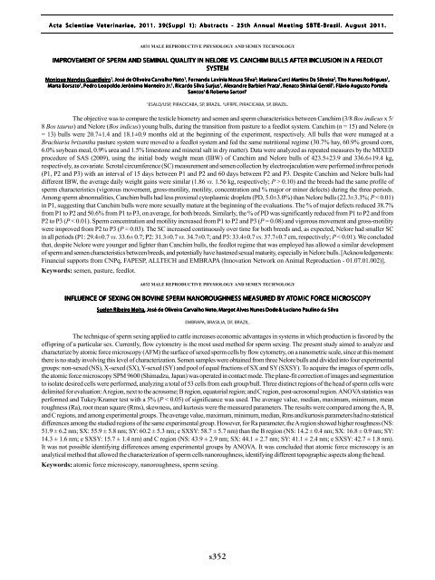
The objective was to compare the testicle biometry and semen and sperm characteristics between Canchim (3/8 Bos indicus x 5/<br />
8 Bos taurus) and Nelore (Bos indicus) young bulls, during the transition from pasture to a feedlot system. Canchim (n = 15) and Nelore (n<br />
= 13) bulls were 20.7±1.4 and 18.1±0.9 months old at the beginning of the experiment, respectively. All bulls that were managed at a<br />
Brachiaria brizantha pasture system were moved to a feedlot system and fed the same nutritional regime (30.7% hay, 60.9% ground corn,<br />
6.0% soybean meal, 0.9% urea and 1.5% limestone and mineral salt in dry matter). Data were analyzed as repeated measures by the MIXED<br />
procedure of SAS (2009), using the initial body weight mean (IBW) of Canchim and Nelore bulls of 423.5±23.9 and 336.6±19.4 kg,<br />
respectively, as covariate. Scrotal circumference (SC) measurement and semen collection by electroejaculation were performed in three periods<br />
(P1, P2 and P3) with an interval of 15 days between P1 and P2 and 60 days between P2 and P3. Despite Canchim and Nelore bulls had<br />
different IBW, the average daily weight gains were similar (1.86 vs. 1.56 kg, respectively; P > 0.10) and the breeds had the same profile of<br />
sperm characteristics (vigorous movement, gross-motility, motility, concentration and % major or minor defects) during the three periods.<br />
Among sperm abnormalities, Canchim bulls had less proximal cytoplasmic droplets (PD, 5.0±3.0%) than Nelore bulls (22.3±3.3%; P < 0.01)<br />
in P1, suggesting that Canchim bulls were more sexually mature at the beginning of the evaluations. The % of major defects reduced 38.7%<br />
from P1 to P2 and 50.6% from P1 to P3, on average, for both breeds. Similarly, the % of PD was significantly reduced from P1 to P2 and from<br />
P2 to P3 (P < 0.01). Sperm concentration and motility increased from P1 to P2 and P3 (P = 0.08) and vigorous movement and gross-motility<br />
were improved from P2 to P3 (P = 0.03). The SC increased continuously over time for both breeds and, as expected, Nelore had smaller SC<br />
in all periods (P1: 29.4±0.7 vs. 33.6± 0.7; P2: 31.3±0.7 vs. 34.7±0.7; and P3: 33.4±0.7 vs. 37.7±0.7 cm, respectively; P < 0.01). We concluded<br />
that, despite Nelore were younger and lighter than Canchim bulls, the feedlot regime that was employed has allowed a similar development<br />
of sperm and semen characteristics between breeds, and potentially have hastened sexual maturity, especially in Nelore bulls. [Acknowledgements:<br />
Financial supports from CNPq, FAPESP, ALLTECH and EMBRAPA (Innovation Network on Animal Reproduction - 01.07.01.002)].<br />
Keywords: semen, pasture, feedlot.<br />
A032 MALE REPRODUCTIVE PHYSIOLOGY AND SEMEN TECHNOLOGY<br />
INFLUENCE OF SEXING ON BOVINE SPERM NANOROUGHNESS OUGHNESS MEASURED BY ATOMIC FORCE MICROSC<br />
OSCOPY<br />
OPY<br />
Suelen Ribeiro Moita, , José de Oliveira Carvalho Neto, Margot Alves Nunes Dode & Luciano Paulino da Silva<br />
EMBRAPA, BRASILIA, DF, BRAZIL.<br />
The technique of sperm sexing applied to cattle increases economic advantages in systems in which production is favored by the<br />
offspring of a particular sex. Currently, flow cytometry is the most used method for sperm sexing. The present study aimed to analyze and<br />
characterize by atomic force microscopy (AFM) the surface of sexed sperm cells by flow cytometry, on a nanometric scale, since at this moment<br />
there is no study involving this level of characterization. Semen samples were obtained from three Nelore bulls and divided into four experimental<br />
groups: non-sexed (NS), X-sexed (SX), Y-sexed (SY) and pool of equal fractions of SX and SY (SXSY). To acquire the images of sperm cells,<br />
the atomic force microscopy SPM 9600 (Shimadzu, Japan) was operated in contact mode. The plane-fit correction of images and segmentation<br />
to isolate desired cells were performed, analyzing a total of 53 cells from each group/bull. Three distinct regions of the head of sperm cells were<br />
delimited for evaluation: A region, next to the acrosome; B region, equatorial region; and C region, post-acrosomal region. ANOVA statistics was<br />
performed and Tukey/Kramer test with a 5% (P < 0.05) of significance was used. The average value, median, maximum, minimum, mean<br />
roughness (Ra), root mean square (Rms), skewness, and kurtosis were the measured parameters. The results were compared among the A, B,<br />
and C regions, and among experimental groups. The average value, maximum, minimum, median, Rms and kurtosis parameters had no statistical<br />
differences among the studied regions of the same experimental group. However, for Ra parameter, the A region showed higher roughness (NS:<br />
51.9 ± 6.2 nm; SX: 55.9 ± 5.8 nm; SY: 60.2 ± 5.3 nm; e SXSY: 58.7 ± 5.7 nm) than the B region (NS: 14.2 ± 0.4 nm; SX: 16.8 ± 0.9 nm; SY:<br />
14.3 ± 1.6 nm; e SXSY: 15.7 ± 1.4 nm) and C region (NS: 43.9 ± 2.9 nm; SX: 44.1 ± 2.7 nm; SY: 41.1 ± 2.4 nm; e SXSY: 42.7 ± 1.8 nm).<br />
It was not possible identifying differences among experimental groups by ANOVA. It was concluded that atomic force microscopy is an<br />
analytical method that allowed the characterization of sperm cells nanoroughness, identifying different topographic aspects along the head.<br />
Keywords: atomic force microscopy, nanoroughness, sperm sexing.<br />
s352


