2011 (SBTE) 25th Annual Meeting Proceedings - International ...
2011 (SBTE) 25th Annual Meeting Proceedings - International ...
2011 (SBTE) 25th Annual Meeting Proceedings - International ...
Create successful ePaper yourself
Turn your PDF publications into a flip-book with our unique Google optimized e-Paper software.
Acta Scientiae Veterinariae, <strong>2011</strong>. 39(Suppl 1): Abstracts - <strong>25th</strong> <strong>Annual</strong> <strong>Meeting</strong> <strong>SBTE</strong>-Brazil. August <strong>2011</strong>.<br />
A203 EMBRYOLOGY, BIOLOGY OF DEVELOPMENT AND PHYSIOLOGY OF REPRODUCTION<br />
SUPPLEMENTATION TION OF MEDIA FOR IN VITRO MATUR<br />
TURATION TION OF OOCYTES OF RABBIT<br />
ABBITS<br />
Tássia Mangetti Gonçalves, , Juliano Rodrigues Sangalli, Marcos Roberto Chiaratti & Flávio Vieira Meirelles<br />
FZEA- USP, PIRASSUNUNGA, SP, BRAZIL.<br />
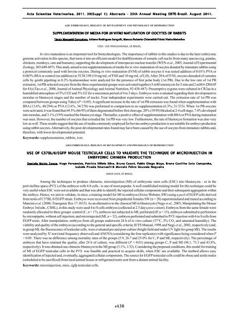
In vitro maturation is an important tool for biotechnologies. The importance of rabbits in this studies is due to the later embryonic<br />
genome activation in this species, that turns it into an efficient model for dedifferentiate of somatic cell nuclei from many species (eg, pandas,<br />
chickens, monkeys, cats and humans), supporting the development of interspecies nuclear transfer (WEN et al., 2005, Journal of Experimental<br />
Zoology, 303:689-697). This study aimed to test supplementations of media for in vitro maturation of oocytes donated by immature rabbits using<br />
a protocol commonly used with bovine oocytes. During in vitro maturation (IVM) of rabbit oocytes it was tested addition of 0.01% PVA or<br />
0.003% BSA or control (no addition) in TCM 199 (10 mg/mL of FSH and 10 mg/mL of LH). After 20 h of IVM, oocytes denuded of cumulus<br />
cells by gentle pipetting in 0.2% hyaluronidase were analyzed for the presence of first polar body (1st PB). Due to the low rate of 1st PB<br />
extrusion, 1st PB-selected oocytes from the three experimental groups were activated together (5 mM ionomycin for 5 min and 2 mM 6-DMAP<br />
for 4 h) (Tao et al., 2008, Journal of Animal Physiology and Animal Nutrition, 92:438-447). Presumptive zygotes were cultured in CR2aa in a<br />
humidified atmosphere of 5% CO2 and 5% O2 for a maximum period of 4 to 5 days. Embryos were evaluated regarding their development to<br />
morulae or blastocyst stages and the number of nuclei. Four independent experiments were carried out. The extrusion rate of 1st PB was<br />
compared between groups using Tukey (P < 0.05). A significant increase in the rate of 1st PB extrusion was found when supplementation with<br />
BSA (13.6%, 40/294) or PVA (12.6%, 34/270) was performed in comparison to no supplementation (6.3%; 21/333). When 1st PB oocytes<br />
were activated, it was found that 69.5% (66/95) of them degenerated before first cleavage, 20% (19/95) blocked at 2-4 cell stage, 7.4% developed<br />
into morulae, and 3.1% (3/95) reached the blastocyst stage. Thereafter, a positive effect of supplementation with BSA or PVA during maturation<br />
was seen. However, the number of oocytes that extruded the 1st PB was very low. Furthermore, the rate of blastocyst formation was also very<br />
low as well. These results suggest that the use of media commonly employed for bovine embryo production is not suitable for embryo production<br />
using rabbit oocytes. Alternatively, the poor developmental rates found may have been caused by the use of oocytes from immature rabbits and,<br />
therefore, with lower developmental potential.<br />
Keywords: supplementation, rabbits, ivm.<br />
A204 EMBRYOLOGY, BIOLOGY OF DEVELOPMENT AND PHYSIOLOGY OF REPRODUCTION<br />
USE OF C57BL/6/EGFP MOUSE TESTICULAR CELLS TO VALIDA<br />
ALIDATE<br />
THE TECHNIQUE OF MICROINJECTION IN<br />
EMBRYONIC CHIMERA PRODUCTION<br />
Daniela Motta Souza, , Hugo Fernandes, Patrícia Villela Silva, Bruno Cazari, Pablo Diego Moço, Bruna Castilho Soto Campanha,<br />
Isabele Picada Emanuelli & Marcelo Fábio Gouveia Nogueira<br />
UNESP, ASSIS, SP, BRAZIL.<br />
Among the techniques to produce chimeras, microinjection (MI) of embryonic stem cells (ESC) into blastocysts - or in the<br />
perivitelline space (PVS.) of the embryos with 4-8 cells - is one of most popular. A well-established training model for this technique could be<br />
very useful when ESC were not available and that was able to identify the injected cellular components and their subsequent aggregation within<br />
the embryo. Hence, we aim to validate, in mice, a training model for MI in embryos (Swiss Webster, SW) using a pool of EGFP cells derived<br />
from testis of C57BL/6/EGFP strain. Embryos were recovered from prepubertal females SW (n = 20) superstimulated and mated according to<br />
Mancini et al. (2008; Transgenic Res 17:1015). As an alternative to the classical MI in blastocysts (Nagy et al., 2003; Manipulating the Mouse<br />
Embryo 3rd edn., CSHL), in this study were used 4 to 8 cells embryos (collected at 2.5 days post coitum). Embryos from the same female were<br />
randomly allocated to three groups: control (C, n = 17), embryos not subjected to MI; perforated (P, n = 15), embryos submitted to perforation<br />
by micropipette, without cell injection; and microinjected (MI, n = 32), embryos perforated and submitted to PVS. injection with 6 to 8 cells from<br />
EGFP testis. After manipulation, embryos from all groups underwent 24 h of in vitro culture (37°C, 5% CO 2<br />
and saturated humidity). The<br />
viability and quality of the embryos (according to the general and specific criteria; IETS Manual, 1998 and Nagy et al., 2003, respectively) and,<br />
in group MI, the fluorescence of testicular cells, were evaluated pre and post-culture (bright field and under UV light for group MI). The results<br />
were analyzed by X 2 test (total frequency observed) and ANOVA (considering the four replicates) with significance being considered when P<br />
< 0.05. There was no difference among mortality rates of the groups (5.9, 26.7 and 25.0% for C, P and MI, respectively). The percentage of<br />
embryos that have retained the quality, after 24 h of culture, was different (P < 0.01) among groups C, P and MI (94.1, 73.3 and 43.8%,<br />
respectively). It was obtained one chimeric blastocyst in the MI group (3.1%, 1/32). Considering the proposed conditions, this model for training<br />
of MI of EGFP testicular cells in the PVS. was feasible and practical to acquire skills, when ESC are available. The method allows easy<br />
identification of injected and, eventually, aggregated cellular components. The source for EGFP testicular cells could be obese and senile males<br />
(scheduled to be sacrificed) from local animal house or refrigerated testis sent from a distant animal facility.<br />
Keywords: microinjection, mice, egfp testicular cells.<br />
s438


