2011 (SBTE) 25th Annual Meeting Proceedings - International ...
2011 (SBTE) 25th Annual Meeting Proceedings - International ...
2011 (SBTE) 25th Annual Meeting Proceedings - International ...
You also want an ePaper? Increase the reach of your titles
YUMPU automatically turns print PDFs into web optimized ePapers that Google loves.
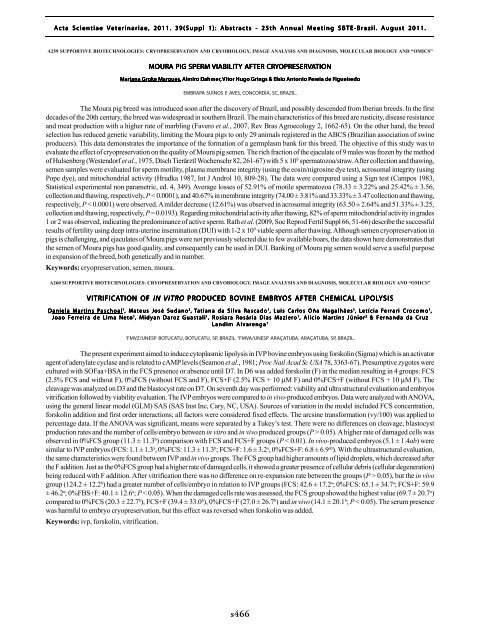
Acta Scientiae Veterinariae, <strong>2011</strong>. 39(Suppl 1): Abstracts - <strong>25th</strong> <strong>Annual</strong> <strong>Meeting</strong> <strong>SBTE</strong>-Brazil. August <strong>2011</strong>.<br />
A259 SUPPORTIVE BIOTECHNOLOGIES: CRYOPRESERVATION AND CRYOBIOLOGY, IMAGE ANALYSIS AND DIAGNOSIS, MOLECULAR BIOLOGY AND “OMICS”<br />
MOURA PIG SPERM VIABILITY AFTER CRYOPRESER<br />
OPRESERVATION<br />
Mar<br />
ariana Grok<br />
oke Mar<br />
arques<br />
ques, Almir<br />
lmiro Dahmer<br />
ahmer, Vit<br />
itor Hugo Grings & Elsio Antonio Per<br />
ereia eia de Figueir<br />
igueiredo<br />
edo<br />
EMBRAPA SUÍNOS E AVES, CONCORDIA, SC, BRAZIL.<br />
The Moura pig breed was introduced soon after the discovery of Brazil, and possibly descended from Iberian breeds. In the first<br />
decades of the 20th century, the breed was widespread in southern Brazil. The main characteristics of this breed are rusticity, disease resistance<br />
and meat production with a higher rate of marbling (Favero et al., 2007, Rev Bras Agroecology 2, 1662-65). On the other hand, the breed<br />
selection has reduced genetic variability, limiting the Moura pigs to only 29 animals registered in the ABCS (Brazilian association of swine<br />
producers). This data demonstrates the importance of the formation of a germplasm bank for this breed. The objective of this study was to<br />
evaluate the effect of cryopreservation on the quality of Moura pig semen. The rich fraction of the ejaculate of 9 males was frozen by the method<br />
of Hulsenberg (Westendorf et al., 1975, Dtsch Tierärztl Wochenschr 82, 261-67) with 5 x 10 9 spermatozoa/straw. After collection and thawing,<br />
semen samples were evaluated for sperm motility, plasma membrane integrity (using the eosin/nigrosine dye test), acrosomal integrity (using<br />
Pope dye), and mitochondrial activity (Hrudka 1987, Int J Androl 10, 809-28). The data were compared using a Sign test (Campos 1983,<br />
Statistical experimental non parametric, ed. 4, 349). Average losses of 52.91% of motile spermatozoa (78.33 ± 3.22% and 25.42% ± 3.56,<br />
collection and thawing, respectively, P < 0.0001), and 40.67% in membrane integrity (74.00 ± 3.81% and 33.33% ± 3.47 collection and thawing,<br />
respectively, P < 0.0001) were observed. A milder decrease (12.61%) was observed in acrosomal integrity (63.50 ± 2.64% and 51.33% ± 3.25,<br />
collection and thawing, respectively, P= 0.0193). Regarding mitochondrial activity after thawing, 82% of sperm mitochondrial activity in grades<br />
1 or 2 was observed, indicating the predominance of active sperm. Rath et al. (2009, Soc Reprod Fertil Suppl 66, 51-66) describe the successful<br />
results of fertility using deep intra-uterine insemination (DUI) with 1-2 x 10 9 viable sperm after thawing. Although semen cryopreservation in<br />
pigs is challenging, and ejaculates of Moura pigs were not previously selected due to few available boars, the data shown here demonstrates that<br />
the semen of Moura pigs has good quality, and consequently can be used in DUI. Banking of Moura pig semen would serve a useful purpose<br />
in expansion of the breed, both genetically and in number.<br />
Keywords: cryopreservation, semen, moura.<br />
A260 SUPPORTIVE BIOTECHNOLOGIES: CRYOPRESERVATION AND CRYOBIOLOGY, IMAGE ANALYSIS AND DIAGNOSIS, MOLECULAR BIOLOGY AND “OMICS”<br />
VITRIFICATION TION OF IN VITRO PRODUCED BOVINE EMBRYOS AFTER CHEMICAL LIPOLYSIS<br />
D aniela Mar<br />
artins Paschoal<br />
1 , Mat eus José Sudano 2 , Tatiana da Silv<br />
ilva Rasc<br />
ascado<br />
ado 1 , Luis Car<br />
arlos Oña Magalhães<br />
1 , Letícia Fer<br />
err ari i Cro c omo 1 ,<br />
Joao Ferreira de Lima Neto 1 , Midyan Daroz Guastali 1 , Rosiara Rosária Dias Maziero 1 , Alicio Martins Júnior 2 & Fernanda da Cruz<br />
Landim Alvarenga 1<br />
1<br />
FMVZ/UNESP BOTUCATU, BOTUCATU, SP, BRAZIL. 2 FMVA/UNESP ARAÇATUBA, ARAÇATUBA, SP, BRAZIL.<br />
The present experiment aimed to induce cytoplasmic lipolysis in IVP bovine embryos using forskolin (Sigma) which is an activator<br />
agent of adenylate cyclase and is related to cAMP levels (Seamon et al., 1981; Proc Natl Acad Sc USA 78, 3363-67). Presumptive zygotes were<br />
cultured with SOFaa+BSA in the FCS presence or absence until D7. In D6 was added forskolin (F) in the median resulting in 4 groups: FCS<br />
(2.5% FCS and without F), 0%FCS (without FCS and F), FCS+F (2.5% FCS + 10 µM F) and 0%FCS+F (without FCS + 10 µM F). The<br />
cleavage was analyzed on D3 and the blastocyst rate on D7. On seventh day was performed: viability and ultra structural evaluation and embryos<br />
vitrification followed by viability evaluation. The IVP embryos were compared to in vivo-produced embryos. Data were analyzed with ANOVA,<br />
using the general linear model (GLM) SAS (SAS Inst Inc, Cary, NC, USA). Sources of variation in the model included FCS concentration,<br />
forskolin addition and first order interactions; all factors were considered fixed effects. The arcsine transformation (vy/100) was applied to<br />
percentage data. If the ANOVA was significant, means were separated by a Tukey’s test. There were no differences on cleavage, blastocyst<br />
production rates and the number of cells/embryo between in vitro and in vivo produced groups (P > 0.05). A higher rate of damaged cells was<br />
observed in 0%FCS group (11.3 ± 11.3 b ) comparison with FCS and FCS+F groups (P < 0.01). In vivo-produced embryos (5.1 ± 1.4ab) were<br />
similar to IVP embryos (FCS: 1.1 ± 1.3 a , 0%FCS: 11.3 ± 11.3 b ; FCS+F: 1.6 ± 3.2 a , 0%FCS+F: 6.8 ± 6.9 ab ). With the ultrastructural evaluation,<br />
the same characteristics were found between IVP and in vivo groups. The FCS group had higher amounts of lipid droplets, which decreased after<br />
the F addition. Just as the 0%FCS group had a higher rate of damaged cells, it showed a greater presence of cellular debris (cellular degeneration)<br />
being reduced with F addition. After vitrification there was no difference on re-expansion rate between the groups (P > 0.05), but the in vivo<br />
group (124.2 ± 12.2 b ) had a greater number of cells/embryo in relation to IVP groups (FCS: 42.6 ± 17.2 a ; 0%FCS: 65.1 ± 34.7 a ; FCS+F: 59.9<br />
± 46.2 a ; 0%FBS+F: 40.1 ± 12.6 a ; P < 0.05). When the damaged cells rate was assessed, the FCS group showed the highest value (69.7 ± 20.7 a )<br />
compared to 0%FCS (20.3 ± 22.7 b ), FCS+F (39.4 ± 33.0 b ), 0%FCS+F (27.0 ± 26.7 b ) and in vivo (14.1 ± 20.1 b ; P < 0.05). The serum presence<br />
was harmful to embryo cryopreservation, but this effect was reversed when forskolin was added.<br />
Keywords: ivp, forskolin, vitrification.<br />
s466


