2011 (SBTE) 25th Annual Meeting Proceedings - International ...
2011 (SBTE) 25th Annual Meeting Proceedings - International ...
2011 (SBTE) 25th Annual Meeting Proceedings - International ...
You also want an ePaper? Increase the reach of your titles
YUMPU automatically turns print PDFs into web optimized ePapers that Google loves.
Acta Scientiae Veterinariae, <strong>2011</strong>. 39(Suppl 1): Abstracts - <strong>25th</strong> <strong>Annual</strong> <strong>Meeting</strong> <strong>SBTE</strong>-Brazil. August <strong>2011</strong>.<br />
A217 CLONING, TRANSGENESIS AND STEM CELLS<br />
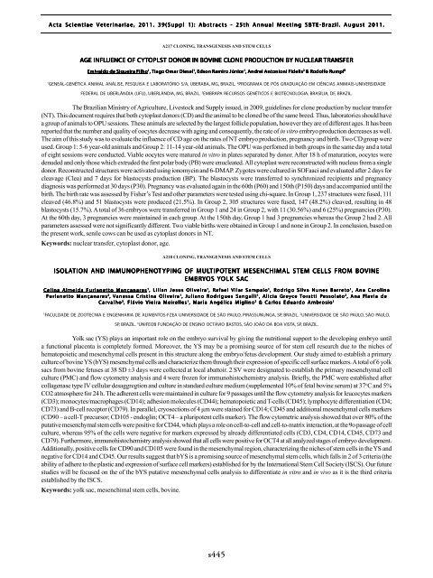
AGE INFLUENCE OF CYTOPLST DONOR IN BOVINE CLONE PRODUCTION BY NUCLEAR TRANSFER<br />
Emivaldo de Siqueira Filho 1 , Tiago Omar Diesel 1 , Edson Ramiro Júnior 1 , Andrei Antonioni Fidelis 2 & Rodolfo Rumpf 3<br />
1<br />
GENEAL-GENÉTICA ANIMAL ANÁLISE, PESQUISA E LABORATÓRIO S/A, UBERABA, MG, BRAZIL. 2 PROGRAMA DE PÓS GRADUAÇÃO EM CIÊNCIAS ANIMAIS-UNIVERSIDADE<br />
FEDERAL DE UBERLÂNDIA (UFU), UBERLÂNDIA, MG, BRAZIL. 3 EMBRAPA RECURSOS GENÉTICOS E BIOTECNOLOGIA, BRASÍLIA, DF, BRAZIL.<br />
The Brazilian Ministry of Agriculture, Livestock and Supply issued, in 2009, guidelines for clone production by nuclear transfer<br />
(NT). This document requires that both cytoplast donors (CD) and the animal to be cloned be of the same breed. Thus, laboratories should have<br />
a group of animals to OPU sessions. These animals are selected by the largest follicle population, however they are of different ages. It has been<br />
reported that the number and quality of oocytes decrease with aging and consequently, the rate of in vitro embryo production decreases as well.<br />
The aim of this study was to evaluate the influence of CD age on the rates of NT embryo production, pregnancy and birth. Two CD group were<br />
used. Group 1: 5-6 year-old animals and Group 2: 11-14 year-old animals. The OPU was perfomed in both groups in the same day and a total<br />
of eight sessions were conducted. Viable oocytes were matured in vitro in plates separated by donor. After 18 h of maturation, oocytes were<br />
denuded and only those which extruded the first polar body (PB) were enucleated. All cytoplast were reconstructed with nucleus from a single<br />
donor. Reconstructed structures were activated using ionomycin and 6-DMAP. Zygotes were cultured in SOFaaci and evaluated after 2 days for<br />
cleavage (Clea) and 7 days for blastocysts production (BP). The blastocysts were transferred to synchronized recipients and pregnancy<br />
diagnosis was performed at 30 days (P30). Pregnancy was evaluated again in the 60th (P60) and 150th (P150) days and accompanied until the<br />
birth. The birth rate was assessed by Fisher’s Test and other parameters were tested using chi-square. In Group 1, 237 structures were fused, 111<br />
cleaved (46.8%) and 51 blastocysts were produced (21.5%). In Group 2, 305 structures were fused, 147 (48.2%) cleaved, resulting in 48<br />
blastocysts (15.7%). A total of 36 embryos were transferred in Group 1 and 24 in Group 2, with 11 (30.56%) and 6 (25%) pregnancies (P30).<br />
At the 60th day, 3 pregnancies were maintained in each group. At the 150th day, Group 1 had 3 pregnancies whereas the Group 2 had 2. All<br />
parameters assessed were not significantly different. Two viable births were obtained in Group 1 and none in Group 2. In conclusion, based on<br />
the present work, senile cows can be used as cytoplast donors in NT.<br />
Keywords: nuclear transfer, cytoplast donor, age.<br />
A218 CLONING, TRANSGENESIS AND STEM CELLS<br />
ISOLATION AND IMMUNOPHENOT YPING OF MULTIPO<br />
TIPOTENT TENT MESENCHIMAL STEM CELLS FROM BOVINE<br />
EMBRYOS<br />
YOLK SAC<br />
Celina Almeida Furlanetto Mançanares 1 , Lilian Jesus Oliveira 1 , Rafael Vilar Sampaio 1 , Rodrigo Silva Nunes Barreto 1 , Ana Carolina<br />
Fur<br />
urlanett<br />
lanetto Mançanar<br />
ançanares<br />
2 , V anessa Cr istina Oliv<br />
liveir<br />
eira 1 , Juliano Ro drigues Sangalli<br />
1 , Alicia Gre y ce Tor<br />
ora tti Pessola<br />
essolato 2 , A na Fla<br />
lavia de<br />
Carvalho 3 , Flávio Vieira Meirelles 1 , Maria Angélica Miglino 2 & Carlos Eduardo Ambrosio 1<br />
1<br />
FACULDADE DE ZOOTECNIA E ENGENHARIA DE ALIMENTOS-FZEA UNIVERSIDADE DE SÃO PAULO, PIRASSUNUNGA, SP, BRAZIL. 2 UNIVERSIDADE DE SÃO PAULO, SÃO PAULO,<br />
SP, BRAZIL. 3 UNIFEOB FUNDAÇÃO DE ENSINO OCTÁVIO BASTOS, SÃO JOÃO DA BOA VISTA, SP, BRAZIL.<br />
Yolk sac (YS) plays an important role on the embryo survival by giving the nutritional support to the developing embryo until<br />
a functional placenta is completely formed. Moreover, the YS may be a promising source of for stem cell research due to the niches of<br />
hematopoietic and mesenchymal cells present in this structure along the embryo/fetus development. Our study aimed to establish a primary<br />
culture of bovine YS (bYS) mesenchymal cells and characterize them through their expression of specific cell surface markers. A total of 6 yolk<br />
sacs from bovine fetuses at 38 SD ±3 days were collected at local abattoir. 2 SV were designated to establish the primary mesenchymal cell<br />
culture (PMC) and flow cytometry analysis and 4 were frozen for immunohistochemistry analysis. Briefly, the PMC were established after<br />
collagenase type IV cellular desaggregtion and culture in standard culture medium (supplemented 10% of fetal bovine serum) at 37 o C and 5%<br />
CO2 atmosphere for 24 h. The adherent cells were maintained in culture for 9 passages until the flow cytometry analysis for leucocytes markers<br />
(CD3); monocytes/macrophages (CD14); adhesion molecules (CD44); hematopoietic and T-cells (CD45); lymphocyte differentiation (CD4;<br />
CD73) and B-cell receptor (CD79). In parallel, cryosections of 4 µm were stained for CD14; CD45 and additional mesenchymal cells markers<br />
(CD90 – a cell-T precursor; CD105 - endoglin; OCT4 – a pluripotent cells marker). The flow cytometric analysis showed that over 80% of the<br />
putative mesenchymal stem cells were positive for CD44, which plays a role on cell-to-cell and cell-to-matrix interaction, at the 9o passage of cell<br />
culture, whereas 95% of the cells were negative for markers expressed by already differentiated cells (CD3, CD4, CD14, CD45, CD73 and<br />
CD79). Furthermore, immunohistochemistry analysis showed that all cells were positive for OCT4 at all analyzed stages of embryo development.<br />
Additionally, positive cells for CD90 and CD105 were found in the mesenchymal region, characterizing the niches of stem cells in the YS and<br />
negative for CD14 and CD45. Our results suggest that bYS is a promising source of mesenchymal stem cells, which falls in 2 of 3 criteria (the<br />
ability of adhere to the plastic and expression of surface cell markers) established for by the <strong>International</strong> Stem Cell Society (ISCS). Our future<br />
studies will be focused on the of the bYS putative mesenchymal cells analysis to differentiate in vitro and in vivo as it is the third criteria<br />
established by the ISCS.<br />
Keywords: yolk sac, mesenchimal stem cells, bovine.<br />
N<br />
s445


