2011 (SBTE) 25th Annual Meeting Proceedings - International ...
2011 (SBTE) 25th Annual Meeting Proceedings - International ...
2011 (SBTE) 25th Annual Meeting Proceedings - International ...
You also want an ePaper? Increase the reach of your titles
YUMPU automatically turns print PDFs into web optimized ePapers that Google loves.
Acta Scientiae Veterinariae, <strong>2011</strong>. 39(Suppl 1): Abstracts - <strong>25th</strong> <strong>Annual</strong> <strong>Meeting</strong> <strong>SBTE</strong>-Brazil. August <strong>2011</strong>.<br />
A253 SUPPORTIVE BIOTECHNOLOGIES: CRYOPRESERVATION AND CRYOBIOLOGY, IMAGE ANALYSIS AND DIAGNOSIS, MOLECULAR BIOLOGY AND “OMICS”<br />
IN VITRO MATUR<br />
TURATION TION OF IMMATURE OOCYTES<br />
VITRIFIED IN DIFFERENT CONDITIONS<br />
Fabiana For<br />
orell<br />
ell, Ner<br />
erissa Albino<br />
lbino, Jamir Machado Junior<br />
unior, Fabiano Car<br />
armina<br />
minatti Zago & Alc<br />
lceu Mezzalir<br />
zzalira<br />
CAV/UDESC, LAGES, SC, BRAZIL.<br />
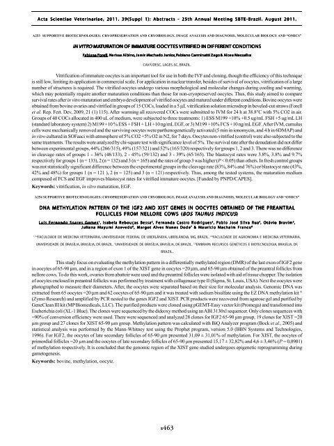
Vitrification of immature oocytes is an important tool for use in both the IVF and cloning, though the efficiency of this technique<br />
is still low, limiting its application in commercial scale. For application in nuclear transfer, besides of survival of oocytes, vitrification of a large<br />
number of structures is required. The vitrified oocytes undergo various morphological and molecular changes during cooling and warming,<br />
which may potentially require another maturation conditions than those for non-cryopreserved oocytes. Thus, this study aimed to compare<br />
survival rates after in vitro maturation and embryo development of vitrified oocytes and matured under different conditions. Bovine oocytes were<br />
obtained from bovine ovaries and vitrified in groups of 15 COCs, loaded in a 5 µL vitrification solution microdrop in beveled-cut straws (Forell<br />
et al. Rep. Fert. Dev, 2009, 21 (1) 115). After warming all recovered COCs were submitted to IVM for 24 h at 38.8°C with 5% CO2 in air.<br />
Groups of 40 COCs allocated in 400 uL of medium, were subjected to three treatments: 1) ESS M199 +10% +0.5 ug/mL FSH +5 ug/mL LH<br />
(standard laboratory system) 2) M199 +10 % ESS + FSH + LH +10 ng/mL EGF, or 3) M199 +10% FCS +10 ng/mL EGF. After IVM, cumulus<br />
cells were mechanically removed and the surviving oocytes were parthenogenetically activated (5 min in ionomycin, and 4 h in 6DMAP) and<br />
in vitro cultured in SOFaaci with atmosphere of 5% CO2 +5% O2 in N2, for 7 days. Oocytes non-vitrified (control) were also subjected to the<br />
same treatments. The results were analyzed by chi-square test with significance level of 5%. The survival rate after the denudation did not differ<br />
between experimental groups, 44% (266/315), 49% (157/321) and 52% (165/320) respectively for groups 1, 2 and 3. There was no difference<br />
in cleavage rates of groups 1 - 36% (48/133), 2 - 45% (59/132) and 3 - 39% (65/165). The blastocyst rates were 3.8%, 3.8% and 9.7%<br />
respectively for groups 1 (n = 133), 2 (n = 132) and 3 (n = 165) and the rates of group 3 was higher (P < 0.05) than others. In fresh control groups<br />
was not statistically significant difference between the experimental groups in the cleavage rate (83%, 84% and 76%) or blastocyst rate (43%,<br />
42% and 48%) for groups 1 (n = 121 ), 2 (n = 125) and 3 (n = 121) respectively. Thus, among the tested systems, the maturation medium<br />
composed of FCS and EGF improves blastocyst rates for vitrified immature oocytes. [Funded by PNPD/CAPES].<br />
Keywords: vitrification, in vitro maturation, EGF.<br />
A254 SUPPORTIVE BIOTECHNOLOGIES: CRYOPRESERVATION AND CRYOBIOLOGY, IMAGE ANALYSIS AND DIAGNOSIS, MOLECULAR BIOLOGY AND “OMICS”<br />
DNA METHYLATION TION PAT TERN OF THE IGF2 AND XIST GENES IN OOCYTES OBTAINED OF THE PREANTRAL<br />
AL<br />
FOLLICLES FROM NELLORE COWS (<br />
(BOS<br />
TAUR<br />
URUS<br />
US INDICUS)<br />
Luís Fernando Soares Gomes 1 , Isabela Rebouças Bessa 2 , Fernanda Castro Rodrigues 3 , Pablo José Silva Rua 4 , Otávio Bravim 5 ,<br />
Juliana Mayumi Azevedo 6 , Margot Alves Nunes Dode 7 & Maurício Machaim Franco 8<br />
1,3,4<br />
FACULDADE DE MEDICINA VETERINÁRIA, UNIVERSIDADE FEDERAL DE UBERLÂNDIA, UBERLÂNDIA, MG, BRAZIL. 2,6 FACULDADE DE AGRONOMIA E MEDICINA VETERINÁRIA,<br />
UNIVERSIDADE DE BRASÍLIA, BRASÍLIA, DF, BRAZIL. 5 UNIVERSIDADE DE BRASÍLIA, BRASÍLIA, DF, BRAZIL. 7,8 EMBRAPA RECURSOS GENÉTICOS E BIOTECNOLOGIA, BRASÍLIA, DF,<br />
BRAZIL.<br />
This study focus on evaluating the methylation pattern in a differentially methylated region (DMR) of the last exon of IGF2 gene<br />
in oocytes of 65-90 µm, and in a region of exon 1 of the XIST gene in oocytes =20 µm, and 65-90 µm obtained of the preantral follicles from<br />
nellore cows. To do this work, ovaries from abattoir were used and the preantral follicles were isolated with aid of tissue chopper. The isolation<br />
of oocytes enclosed in preantral follicles was performed by treatment with collagenase type II (Sigma, St. Louis, USA). Next the oocytes were<br />
photographed to measure their diameters. After, the oocytes were separated based on their size for molecular analysis. Genomic DNA was<br />
extracted from 65 oocytes =20 µm and 62 oocytes of 65-90 µm and it was treated with sodium bisulfate using the EZ DNA methylation kit ®<br />
(Zymo Research) and amplified by PCR nested to the genes IGF2 and XIST. PCR products were recovered from agarose gel and purified by<br />
GeneClean III kit (MP Biomedicals, LLC). The purified products were cloned using pGEMT-Easy vector kit (Promega) and transformed into<br />
Escherichia coli (XL-1 Blue). The clones were sequenced by the dideoxy method using an ABI 3130xl sequencer. Only clones sequences with<br />
=90% of conversion efficiency were used. There were sequenced and analyzed 28 clones for IGF2 65-90 µm group, 19 clones for XIST =20<br />
µm group and 27 clones for XIST 65-90 µm group. Methylation pattern was calculated with BiQ Analyzer program (Bock et al., 2005) and<br />
statistical analysis was performed by the Mann-Whitney test using the Prophet program, version 5.0 (BBN Systems and Technologies,<br />
1996). For IGF2, the oocytes of late secondary follicles of 65-90 µm presented 31,09 ± 31,01% of methylation. For XIST, the oocytes of<br />
primordial follicles =20 µm and the oocytes of late secondary follicles of 65-90 µm presented 15,17 ± 32,82% and 4,6 ± 3,46% (P = 0,0901)<br />
of methylation respectively. It is concluded that the genomic region of the XIST gene studied undergoes epigenetic reprogramming during<br />
gametogenesis.<br />
Keywords: bovine, methylation, oocyte.<br />
N<br />
s463


