2011 (SBTE) 25th Annual Meeting Proceedings - International ...
2011 (SBTE) 25th Annual Meeting Proceedings - International ...
2011 (SBTE) 25th Annual Meeting Proceedings - International ...
Create successful ePaper yourself
Turn your PDF publications into a flip-book with our unique Google optimized e-Paper software.
Acta Scientiae Veterinariae, <strong>2011</strong>. 39(Suppl 1): Abstracts - <strong>25th</strong> <strong>Annual</strong> <strong>Meeting</strong> <strong>SBTE</strong>-Brazil. August <strong>2011</strong>.<br />
A235 SUPPORTIVE BIOTECHNOLOGIES: CRYOPRESERVATION AND CRYOBIOLOGY, IMAGE ANALYSIS AND DIAGNOSIS, MOLECULAR BIOLOGY AND “OMICS”<br />
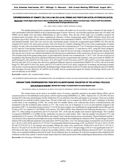
CRYOPRESER<br />
OPRESERVATION OF SOMATIC CELLS IN 0.5 ML OR 0.25 ML STRAWS S AND PROPYLENE GLYCOL OR ETHYLENE GLYCOL<br />
Monica Urio 1 , Claudia Regina Appio Duarte 1 , Renata Cristina Fleith 1 , Maria Luiza Munhoz 1 , Fabiano Carminatti Zago 1 , Fabiana Forell 1 Alceu Mezzalira 1 ,<br />
Marcelo Bertolini 2 & Ubirajara Maciel da Costa 1<br />
1<br />
CAV - UDESC, LAGES, SC, BRAZIL. 2 UNIFOR, FORTALEZA, CE, BRAZIL.<br />
The standard protocol for the cryopreservation of somatic cells makes use of cryovials as classic containers for the storage of<br />
cells, and dimethyl sulfoxide (DMSO) as the cryoprotectant agent of choice. However, cryovials take significant space into LN2 tanks, and<br />
DMSO can be highly toxic and induce differentiation in cells in culture. Thus, the aim of this study was to establish a protocol for<br />
cryopreservation of somatic cells in straws comparing the efficiency of three cryoprotectant agents: DMSO, Ethylene Glycol (EG) and<br />
Propylene Glycol (PG). Primary cell cultures of bovine fibroblasts were maintained in DMEM + 10% FCS in an incubator at 5% CO2 in air<br />
and saturated humidity. Such cells were used for nine experimental groups varying the cryoprotectant agent (DMSO, EG, PG) and the<br />
container (0.25 and 0.5 mL straws and cryovials). Cells were frozen in a solution containing 10% of one of the cryoprotectants in culture<br />
medium. For this, cells were loaded into the container and exposed to the cryoprotectant at 4° C for 15 min before freezing. Straws were kept<br />
in LN2 vapor for 5 min and then immersed in LN2, whereas cryovials were cooled at -1°C/min down to -80°C, using Mr. Frosty ® (Nalgene),<br />
and then transferred to LN2. The experiment was separated in two steps for the survival study, evaluated by the Trypan blue staining. In the<br />
first step, the survival rate was evaluated immediately after thawing (seven replications). In the second step, cell survival was evaluated after<br />
24 h of culture (only for the groups usning straws, in nine replications). Results were evaluated by the ÷2, for P < 0.05. The rate of cell survival<br />
immediately after thawing showed no significant differences between the cryoprotectants and containers, with survival rates for DMSO, EG<br />
and PG being 83%, 86% and 87% in 0.5 mL straws ; 81%, 83% and 87% in 0,25 mL straws; and 87%, 90% and 91% in cryovials, respectively.<br />
Survival rates after 24 h of culture also showed no significant difference between cryoprotectants (DMSO, EG or PG), with survival rates<br />
being 91%, 90% and 91% for 0.5 mL straws, and 91%, 91% and 93% for 0.25 mL straws, respectively. In summary, the freezing of somatic<br />
cells in straws was proven practical and feasible, facilitating storage of larger batches of cells in LN2 tanks, also providing the same cell viability<br />
after freezingas cryovials. Moreover, EG and PG can be readily used as cryoprotectant agents for somatic cell freezing with the same cell<br />
survival efficiency as for DMSO.<br />
Keywords: somatic cell, cryoprotectants, straws.<br />
A236 SUPPORTIVE BIOTECHNOLOGIES: CRYOPRESERVATION AND CRYOBIOLOGY, IMAGE ANALYSIS AND DIAGNOSIS, MOLECULAR BIOLOGY AND “OMICS”<br />
OVARIAN<br />
TISSUE CRYOPRESER<br />
OPRESERVATION FROM BITCHES:<br />
MORPHOLOGIC OGIC EVAL<br />
ALUATION OF PRE-ANTRALS ALS FOLLICLES<br />
José Luiz Jivago de Paula Rôlo 1 , Michelle Silva Araújo 2 , Fernanda Paulini 1 & Carolina Madeira Lucci 1<br />
1<br />
PPG BIOLOGIA ANIMAL, IB, UNB, BRASILIA, DF, BRAZIL. 2 FACULDADE DE AGRONOMIA E MEDICINA VETERINÁRIA, UNB, BRASILIA, DF, BRAZIL.<br />
The ovarian tissue can be used as an storable source of oocytes, especially enclosed in pre-antral follicles (PAF), and its<br />
cryopreservation is well established in some species, with few reports in dogs (ISHIJIMA et al., 2006, J. Reprod. Dev. 52, 293-299). The slow<br />
freezing (SF) is the most used technique, but the vitrification (V) is an alternative that offers advantages in convenience. There are no studies<br />
comparing the two methods in bitch ovarian tissue. The aim of this study was to compare the effects of SF and V techniques in the bitch<br />
ovarian tissue in the morphology of PAF. Ten ovaries from five healthy bitches were collected after elective ovariohysterectomy and<br />
transported to the lab in buffered saline solution at 37°C. The ovaries were cut in fragments of 1x1x3 cm and randomly assigned to the<br />
procedures of SF, V or immediate fixation (control, C). For SF, the tissue samples were equilibrated in a programmable freezer (Biocom) for<br />
20 min at 10°C in a solution of 1.5M DMSO, 0.4% sucrose, 10% FBS in MEM. Then submitted to the following freezing curve: -1°C/min<br />
until -7°C, when seeding was performed, and -0.3°C/min until -30°C. At the end, the samples were stored in liquid nitrogen. The V method<br />
was based on ISHIJIMA et al. (2006), but the vitrification was done in a solid surface. After 7 days the samples were placed at 37°C and<br />
equilibrated in 0.4% sucrose, 10% FBS and MEM, and washed 3 times with decreasing sucrose concentration. Thawed fragments and the C<br />
were fixed in Carnoy and processed for light microscopy with HE staining for the morphological evaluation of PAF. The percentages of normal<br />
follicles (NF) in the different treatments were analyzed by ANOVA and Tukey´s test. The total percentage of normal PAF was 93.66±6.81%<br />
for C, 86.16±11.05% for SF and 68.14±12.75% for V. For primordial follicles the percentage of NF was 96.69±4.72% for C, 89.51±10.39%<br />
SF and 75.32±9.23% in V. For primary and secondary follicles the values were, respectively, 94.80±6.91% and 87.62±17.12% for C,<br />
86.8±12.15% and 76.35±26.34% for SF and 61.53±14.78 and 52.25±22.13% for V. The percentage of NF in V was significantly lower (P <<br />
0.05) than in the other two treatments, except for the secondary follicles, where no significant difference between SF and V was observed.<br />
There was no significant difference between SF and C for any follicular class. In conclusion, SF is the best cryopreservation method for PAF<br />
in bitches ovarian tissue, and the V protocol needs improvement for a better preservation of the morphological characteristics of the follicles.<br />
Keywords: slow freezing, vitrification, oocyte.<br />
s454


