2011 (SBTE) 25th Annual Meeting Proceedings - International ...
2011 (SBTE) 25th Annual Meeting Proceedings - International ...
2011 (SBTE) 25th Annual Meeting Proceedings - International ...
You also want an ePaper? Increase the reach of your titles
YUMPU automatically turns print PDFs into web optimized ePapers that Google loves.
Acta Scientiae Veterinariae, <strong>2011</strong>. 39(Suppl 1): Abstracts - <strong>25th</strong> <strong>Annual</strong> <strong>Meeting</strong> <strong>SBTE</strong>-Brazil. August <strong>2011</strong>.<br />
A211 CLONING, TRANSGENESIS AND STEM CELLS<br />
EFFECT OF TRICHOSTAIN A ON PRODUCTION OF BOVINE EMBRYOS PRODUCED BY SOMATIC CELL NUCLEAR TRANSFER<br />
AND SUBSEQUENT GESTATIONS<br />
TIONS<br />
Luiz Sérgio Almeida Camar<br />
amargo<br />
go, Car<br />
arolina Cap<br />
apobiango R. Quin<br />
uintão<br />
tão, Anna Car<br />
arolina Denic<br />
enicol,<br />
Lilian Tam<br />
amy Iguma, Bruno Camp<br />
ampos Car<br />
aravalho<br />
alho, Luiz Gusta<br />
ustavo Bruno<br />
Siqueira & João Henrique Moreira Viana<br />
EMBRAPA GADO DE LEITE, JUIZ DE FORA, MG, BRAZIL.<br />
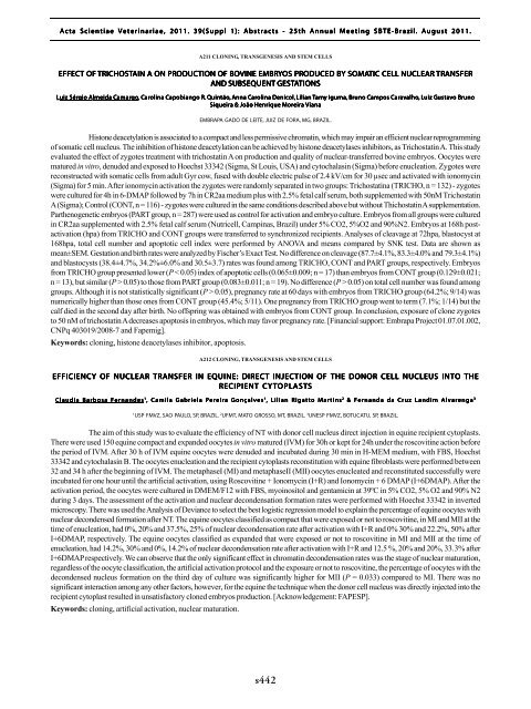
Histone deacetylation is associated to a compact and less permissive chromatin, which may impair an efficient nuclear reprogramming<br />
of somatic cell nucleus. The inhibition of histone deacetylation can be achieved by histone deacetylases inhibitors, as Trichostatin A. This study<br />
evaluated the effect of zygotes treatment with trichostatin A on production and quality of nuclear-transferred bovine embryos. Oocytes were<br />
matured in vitro, denuded and exposed to Hoechst 33342 (Sigma, St Louis, USA) and cytochalasin (Sigma) before enucleation. Zygotes were<br />
reconstructed with somatic cells from adult Gyr cow, fused with double electric pulse of 2.4 kV/cm for 30 µsec and activated with ionomycin<br />
(Sigma) for 5 min. After ionomycin activation the zygotes were randomly separated in two groups: Trichostatina (TRICHO, n = 132) - zygotes<br />
were cultured for 4h in 6-DMAP followed by 7h in CR2aa medium plus with 2.5% fetal calf serum, both supplemented with 50nM Trichostatin<br />
A (Sigma); Control (CONT, n = 116) - zygotes were cultured in the same conditions described above but without Thichostatin A supplementation.<br />
Parthenogenetic embryos (PART group, n = 287) were used as control for activation and embryo culture. Embryos from all groups were cultured<br />
in CR2aa supplemented with 2.5% fetal calf serum (Nutricell, Campinas, Brazil) under 5% CO2, 5%O2 and 90%N2. Embryos at 168h postactivation<br />
(hpa) from TRICHO and CONT groups were transferred to synchronized recipients. Analyses of cleavage at 72hpa, blastocyst at<br />
168hpa, total cell number and apoptotic cell index were performed by ANOVA and means compared by SNK test. Data are shown as<br />
mean±SEM. Gestation and birth rates were analyzed by Fischer’s Exact Test. No difference on cleavage (87.7±4.1%, 83.3±4.0% and 79.3±4.1%)<br />
and blastocysts (38.4±4.7%, 34.2%±6.0% and 30.5±3.7) rates was found among TRICHO, CONT and PART groups, respectively. Embryos<br />
from TRICHO group presented lower (P < 0.05) index of apoptotic cells (0.065±0.009; n = 17) than embryos from CONT group (0.129±0.021;<br />
n = 13), but similar (P > 0.05) to those from PART group (0.083±0.011; n = 19). No difference (P > 0.05) on total cell number was found among<br />
groups. Although it is not statistically significant (P > 0.05), pregnancy rate at 60 days with embryos from TRICHO group (64.2%; 9/14) was<br />
numerically higher than those ones from CONT group (45.4%; 5/11). One pregnancy from TRICHO group went to term (7.1%; 1/14) but the<br />
calf died in the second day after birth. No offspring was obtained with embryos from CONT group. In conclusion, exposure of clone zygotes<br />
to 50 nM of trichostatin A decreases apoptosis in embryos, which may favor pregnancy rate. [Financial support: Embrapa Project 01.07.01.002,<br />
CNPq 403019/2008-7 and Fapemig].<br />
Keywords: cloning, histone deacetylases inhibitor, apoptosis.<br />
A212 CLONING, TRANSGENESIS AND STEM CELLS<br />
EFFICIENCY OF NUCLEAR TRANSFER IN EQUINE: DIRECT INJECTION OF THE DONOR CELL NUCLEUS INTO THE<br />
RECIPIENT CYTOPL<br />
OPLAST<br />
ASTS<br />
Claudia Barbosa Fernandes 1 , Camila Gabriela Pereira Gonçalves 1 , Lilian Rigatto Martins 2 & Fernanda da Cruz Landim Alvarenga 3<br />
1<br />
USP FMVZ, SAO PAULO, SP, BRAZIL. 2 UFMT, MATO GROSSO, MT, BRAZIL. 3 UNESP FMVZ, BOTUCATU, SP, BRAZIL.<br />
The aim of this study was to evaluate the efficiency of NT with donor cell nucleus direct injection in equine recipient cytoplasts.<br />
There were used 150 equine compact and expanded oocytes in vitro matured (IVM) for 30h or kept for 24h under the roscovitine action before<br />
the period of IVM. After 30 h of IVM equine oocytes were denuded and incubated during 30 min in H-MEM medium, with FBS, Hoechst<br />
33342 and cytochalasin B. The oocytes enucleation and the recipient cytoplasts reconstitution with equine fibroblasts were performed between<br />
32 and 34 h after the beginning of IVM. The metaphaseI (MI) and metaphaseII (MII) oocytes enucleated and reconstituted successfully were<br />
incubated for one hour until the artificial activation, using Roscovitine + Ionomycin (I+R) and Ionomycin + 6 DMAP (I+6DMAP). After the<br />
activation period, the oocytes were cultured in DMEM/F12 with FBS, myoinositol and gentamicin at 39ºC in 5% CO2, 5% O2 and 90% N2<br />
during 3 days. The assessment of the activation and nuclear decondensation formation rates were performed with Hoechst 33342 in inverted<br />
microscopy. There was used the Analysis of Deviance to select the best logistic regression model to explain the percentage of equine oocytes with<br />
nuclear decondensed formation after NT. The equine oocytes classified as compact that were exposed or not to roscovitine, in MI and MII at the<br />
time of enucleation, had 0%, 20% and 37.5%, 25% of nuclear decondensation rate after activation with I+R and 0% 30% and 22.2%, 50% after<br />
I+6DMAP, respectively. The equine oocytes classified as expanded that were exposed or not to roscovitine in MI and MII at the time of<br />
enucleation, had 14.2%, 30% and 0%, 14.2% of nuclear decondensation rate after activation with I+R and 12.5 %, 20% and 20%, 33.3% after<br />
I+6DMAP respectively. We can observe that the only significant effect in chromatin decondensation rates was the stage of nuclear maturation,<br />
regardless of the oocyte classification, the artificial activation protocol and the exposure or not to roscovitine, the percentage of oocytes with the<br />
decondensed nucleus formation on the third day of culture was significantly higher for MII (P = 0.033) compared to MI. There was no<br />
significant interaction among any other factors, however, for the equine the technique when the donor cell nucleus was directly injected into the<br />
recipient cytoplast resulted in unsatisfactory cloned embryos production. [Acknowledgement: FAPESP].<br />
Keywords: cloning, artificial activation, nuclear maturation.<br />
s442


