EXPLORATIONS IN TURKESTAN
EXPLORATIONS IN TURKESTAN
EXPLORATIONS IN TURKESTAN

Create successful ePaper yourself
Turn your PDF publications into a flip-book with our unique Google optimized e-Paper software.
HUMAN REMA<strong>IN</strong>S FROM THE NORTH KURGAN.<br />
457<br />
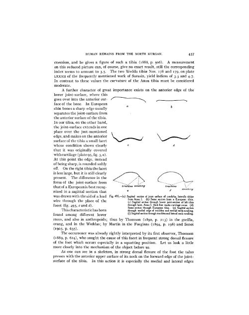
cnemism, and he gives a figure of such a tibia (X888, p. 506). A measurement<br />
on this reduced picture can, of course, give no exact result, still the corresponding<br />
index seems to amount to 3.5. The two Wedda tibiae Nos. 178 and 179, on plate<br />
LXXXII of the frequently mentioned work of Sarasin, yield indices of 3.5 and 4.7.<br />
In contrast to these values the curvature of the Anau tibia must be considered<br />
moderate.<br />
A further character of great importance exists on the anterior edge of the<br />
lower joint-surface, where this<br />
goes over into the anterior sur- -"-- -__<br />
face of the bone. In European<br />
shin bones a sharp edge usually<br />
separates the joint-surface from<br />
the anterior surface of the tibia.<br />
In our tibia, on the other hand,<br />
the joint-surface extends in one<br />
place over the just-mentioned<br />
edge, and makes on the anterior<br />
surface of the tibia a small facet<br />
whose condition shows clearly c d<br />
that it was originally covered<br />
with cartilage (plate 95, fig. 3, a).<br />
At this point the edge, instead<br />
of being sharp, is rounded softly<br />
off. On the right tibia the facet<br />
is less large, but it is still clearly<br />
present. The difference in the .\/<br />
.<br />
form of the joint-surface from , -.. . --<br />
that of a European is best recog- trochtea sweLiny trochlea<br />
nized in a sagittal section that e f<br />
was drawn with the aid of a lead Fig. 495.-(a) Sagittal section of joint surface of condylus lateralis tibiae<br />
,wire through the place of the<br />
w ~ire the through place ~of the<br />
from Anau 1. (b) Same section from a European tibia.<br />
(c) Sagittal section through lower joint-section of left tibia<br />
facet (fig. 495, C and d). through facet, Anau I; thick line marks cartilage cover. (d)<br />
This characteristic has been<br />
found among different lower<br />
Same section through European tibia. (e) Sagittal section<br />
through medial edge of trochlea and medial neck-swelling.<br />
(f) Sagittal section through trochlea and lateral neck-swelling.<br />
races, and also in anthropoids; thus by Thomson (I890, p. 213) in the gorilla,<br />
orang, and in the Weddas; by Martin in the Fuegians (1894, p. 198) and Senoi<br />
(1905, p. 635).<br />
The occurrence was already rightly interpreted by its first observer, Thomson<br />
(1889, p. 624), who sought the cause of this facet in frequent strong dorsal flexure<br />
of the foot which occurs especially in a squatting position. Let us look a little<br />
more closely into the mechanism of the object before us.<br />
As one can see in a skeleton, in strong dorsal flexure of the foot the talus<br />
presses with the anterior upper surface of its neck on the forward edge of the jointsurface<br />
of the tibia. In this action it is especially the medial and lateral edges