EXPLORATIONS IN TURKESTAN
EXPLORATIONS IN TURKESTAN
EXPLORATIONS IN TURKESTAN
You also want an ePaper? Increase the reach of your titles
YUMPU automatically turns print PDFs into web optimized ePapers that Google loves.
458<br />
HUMAN REMA<strong>IN</strong>S FROM THE NORTH KURGAN.<br />
of the neck which are subjected to the pressure, while the central depressed part<br />
is less affected. Accordingly the cartilaginous covering of the joint is spread<br />
especially over the two lateral swellings of the neck; the depression between these<br />
remains free (plate 95, fig. 3, b). But now the two swellings act very differently.<br />
The medial swelling presents a direct continuation of the medial edge of the surface<br />
of the trochlea, as we can see by taking a corresponding impression with<br />
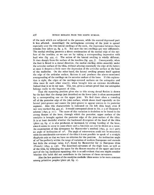
lead wire (fig. 495, e). The action of the lateral swelling is wholly different.<br />
It rises sharply from the surface of the trochlea (fig. 495, f). Consequently, when<br />
the foot is flexed in a dorsal direction, the medial swelling slides smoothly under<br />
the articular surface of the tibia, without altering essentially the edge of the latter;<br />
at most it deepens a little more the depression of the articular surface at the base<br />
of the malleolus. On the other hand, the lateral swelling presses itself against<br />
the edge of the articular surface, flattens it and produces the above-mentioned<br />
overspreading of the cartilage on the anterior surface of the bone. If this explanation<br />
is right, the edges of the cartilage-covered surfaces on the astragalus and<br />
tibia must fit each other exactly, when brought into an extreme dorsiflexion.<br />
Indeed that is so in our case. This, too, gives a certain proof that our astragalus<br />
belongs really to the fragment of tibia.<br />
That the squatting position gives rise to this strong dorsal flexion is shown<br />
by the fact that the change just described on the lower joint is often accompanied<br />
by a corresponding one on the upper joint. We find there often a rounding<br />
off of the posterior edge of the joint-surface, which shows itself especially on the<br />
lateral joint-groove and causes the joint-groove to appear convex in its posterior<br />
segment. Also this characteristic is indicated on the left tibia head, even if<br />
not very marked (fig. 495, a). It would correspond about to No. 2-3 of Thomson's<br />
scheme (1890, p. 21 i). The cause of this variation is doubtless to be found in the<br />
strong flexure of the knee, through which the posterior, upper surface of the<br />
condyles is brought against the posterior edge of the joint-surface of the tibia.<br />
It is at least doubtful whether the backward divergence of the head of the tibia<br />
(plate 95, fig. 2) is also produced, or increased, by strong bending of the knee,<br />
since it seems to occur in cases where such a function can not be shown as a cause.<br />
An examination of this divergence by Manouvrier's method (1893, p. 231) gave<br />
an angle of inclination of o0°. The angle of retroversion could not be measured;<br />
with the considerable curvature of the tibia it is not possible to speak of a straight<br />
diaphysis axis so that we have no criterion for the position. An inclination angle<br />
of 10° lies perfectly within the range of variation of modem Europeans and exceeds<br />
but little the average value, 8.5°, found by Manouvrier for 72 European tibiae<br />
(French) (i893, p. 236). The described curvature of the thigh bone, as well as<br />
of the tibia, by enlarging the space for the flexure muscles of the upper and lower<br />
part of the leg, facilitates squatting; this is so self-evident that one might consider<br />
whether this habit did not contribute to the increase of those curvatures.<br />
Also the low position of the condylus medialis tibiae seems to be more common<br />
among primitive peoples (plate 96, fig. i).


