- Page 1 and 2:
Life Science CATALOG 2013 Table of
- Page 3 and 4:
Cell Signaling Table of Contents Pr
- Page 5 and 6:
Cell Signaling 1 Biobanking 1 DNA E
- Page 7 and 8:
Cell Signaling DNA Extraction for B
- Page 9 and 10:
Cell Signaling Wizard ® Genomic DN
- Page 11 and 12:
Cell Signaling QuantiFluor RNA Syst
- Page 13 and 14:
Cell Signaling Sample ID and Mixed
- Page 15 and 16:
Cell Signaling 2 Biochemicals and L
- Page 17 and 18:
Cell Signaling Agarose, LMP, Prepar
- Page 19 and 20:
Cell Signaling Blue/Orange Loading
- Page 21 and 22:
Cell Signaling Glycine, Molecular B
- Page 23 and 24:
Cell Signaling SDS Solution, Molecu
- Page 25 and 26:
Cell Signaling Tris Base, Molecular
- Page 27 and 28:
Cell Signaling Unmethylated Lambda
- Page 29 and 30:
Cell Signaling Promega Barrier Tips
- Page 31 and 32:
Cell Signaling Magnetic Stands and
- Page 33 and 34:
Cell Signaling 3 Cell Health Assays
- Page 35 and 36:
Cell Signaling A. B. Luminescence/p
- Page 37 and 38:
Cell Signaling Luminogenic Enzyme S
- Page 39 and 40:
Cell Signaling Apoptosis
- Page 41 and 42:
Cell Signaling Caspase-Glo ® 2 Ass
- Page 43 and 44:
Cell Signaling Caspase-Glo ® 8 Ass
- Page 45 and 46:
Cell Signaling CaspACE Assay System
- Page 47 and 48:
Cell Signaling Anti-ACTIVE ® Caspa
- Page 49 and 50:
Cell Signaling CellTiter-Glo ® One
- Page 51 and 52:
Cell Signaling Fluorescent Cell Via
- Page 53 and 54:
Cell Signaling CellTiter-Blue ® Ce
- Page 55 and 56:
Cell Signaling ApoTox-Glo Triplex A
- Page 57 and 58:
Cell Signaling CytoTox-Fluor Cytoto
- Page 59 and 60:
Cell Signaling GSH-Glo Glutathione
- Page 61 and 62:
Cell Signaling 4 Cell Signaling 4 G
- Page 63 and 64:
Cell Signaling cAMP-Glo Max Assay P
- Page 65 and 66:
Cell Signaling Growth Factors Epide
- Page 67 and 68:
Cell Signaling SIRT-Glo Assays and
- Page 69 and 70:
Cell Signaling ADP-Glo Kinase Assay
- Page 71 and 72:
Cell Signaling Kinase Enzyme System
- Page 73 and 74:
Cell Signaling Kinase Enzyme System
- Page 75 and 76:
Cell Signaling Kinase Enzyme System
- Page 77 and 78:
Cell Signaling Kinase Enzyme System
- Page 79 and 80:
Cell Signaling Kinase Enzyme System
- Page 81 and 82:
Cell Signaling ProFluor ® PKA Assa
- Page 83 and 84:
Cell Signaling PepTag ® Non-Radioa
- Page 85 and 86:
Cell Signaling Product Size Cat.# P
- Page 87 and 88:
Cell Signaling kDa 98 - 64 - 50 - 3
- Page 89 and 90:
Cell Signaling ProFluor ® Ser/Thr
- Page 91 and 92:
Cell Signaling 5 Cloning and DNA Ma
- Page 93 and 94:
Cell Signaling DNA Step Ladders Pro
- Page 95 and 96:
Cell Signaling Conventional DNA Mar
- Page 97 and 98:
Cell Signaling RNA Markers Product
- Page 99 and 100:
Cell Signaling AatII 37 Product Siz
- Page 101 and 102:
Cell Signaling BstEII 60 Product Si
- Page 103 and 104:
Cell Signaling HhaI 37 Product Size
- Page 105 and 106:
Cell Signaling NdeI 37 Product Size
- Page 107 and 108:
Cell Signaling SnaBI 37 Product Siz
- Page 109 and 110:
Cell Signaling Alkaline Phosphatase
- Page 111 and 112:
Cell Signaling T4 DNA Polymerase Pr
- Page 113 and 114:
Cell Signaling Kinases and DNA Labe
- Page 115 and 116:
Cell Signaling Additional Enzymes S
- Page 117 and 118:
Cell Signaling Subcloning Tools and
- Page 119 and 120:
Cell Signaling pALTER ® -MAX Vecto
- Page 121 and 122:
Cell Signaling pGEM ® -7Zf(+/-) Ve
- Page 123 and 124:
Cell Signaling pSP72 Vector Product
- Page 125 and 126:
Cell Signaling 6 DNA and RNA Purifi
- Page 127 and 128:
Cell Signaling x-tracta Gel Extract
- Page 129 and 130:
Cell Signaling Wizard ® MagneSil
- Page 131 and 132:
Cell Signaling ReliaPrep Blood gDNA
- Page 133 and 134:
Cell Signaling Wizard ® SV 96 Geno
- Page 135 and 136:
Cell Signaling Fixed-Tissue Genomic
- Page 137 and 138:
Cell Signaling Maxwell ® 16 System
- Page 139 and 140:
Cell Signaling PureYield Plasmid Mi
- Page 141 and 142:
Cell Signaling Wizard ® Plus Minip
- Page 143 and 144:
Cell Signaling Wizard ® SV 96 and
- Page 145 and 146:
Cell Signaling RNA Purification Rel
- Page 147 and 148:
Cell Signaling PureYield RNA Midipr
- Page 149 and 150:
Cell Signaling Maxwell ® 16 System
- Page 151 and 152: Cell Signaling RNasin ® Plus RNase
- Page 153 and 154: Cell Signaling DNA and RNA Quantita
- Page 155 and 156: Cell Signaling Magnetic Stands and
- Page 157 and 158: Cell Signaling Vacuum Manifolds and
- Page 159 and 160: Cell Signaling 7 Drug Discovery 7 D
- Page 161 and 162: Cell Signaling GPCR Assays cAMP-Glo
- Page 163 and 164: Cell Signaling GloResponse Lucifera
- Page 165 and 166: Cell Signaling DPPIV-Glo Protease A
- Page 167 and 168: Cell Signaling Calpain-Glo Protease
- Page 169 and 170: Cell Signaling 8 Epigenetics 8 DNA
- Page 171 and 172: Cell Signaling DNA Purification Tec
- Page 173 and 174: Cell Signaling Luciferase-Based Met
- Page 175 and 176: Cell Signaling GoTaq ® Long PCR Ma
- Page 177 and 178: Cell Signaling DUB-Glo Protease Ass
- Page 179 and 180: Cell Signaling HaloTag ® Protein P
- Page 181 and 182: Cell Signaling 9 Genetic Identity 9
- Page 183 and 184: Cell Signaling DNA IQ System Produc
- Page 185 and 186: Cell Signaling DNA IQ Casework Pro
- Page 187 and 188: Cell Signaling STR Analysis for For
- Page 189 and 190: Cell Signaling PowerPlex ® Y23 Sys
- Page 191 and 192: Cell Signaling PowerPlex ® 18D Sys
- Page 193 and 194: Cell Signaling PowerPlex ® S5 Syst
- Page 195 and 196: Cell Signaling GenePrint ® Fluores
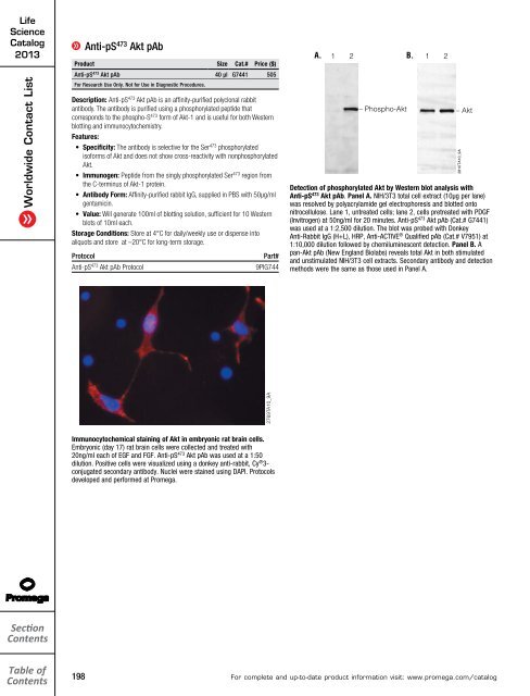
- Page 197 and 198: Cell Signaling 10 Imaging and Immun
- Page 199 and 200: Cell Signaling HaloTag ® Ligand Bu
- Page 201: Cell Signaling NGF E max ® ImmunoA
- Page 205 and 206: Cell Signaling A. Anti-ACTIVE ® MA
- Page 207 and 208: Cell Signaling Anti-ACTIVE ® Caspa
- Page 209 and 210: Cell Signaling Anti-NGF mAb Product
- Page 211 and 212: Cell Signaling Anti-TGFβ 1 pAb Pro
- Page 213 and 214: Cell Signaling Horseradish Peroxida
- Page 215 and 216: Cell Signaling ViviRen In Vivo Reni
- Page 217 and 218: Cell Signaling 11 Industrial and En
- Page 219 and 220: Cell Signaling QuantiLum ® Recombi
- Page 221 and 222: 12 Instruments 12 Luminometers 218
- Page 223 and 224: GloMax ® 20/20 Luminometer Product
- Page 225 and 226: Fluorescence Module: Application-op
- Page 227 and 228: GloMax ® -Multi Jr Single-Tube Mul
- Page 229 and 230: Maxwell ® CSC System for IVD Use M
- Page 231 and 232: Maxwell ® Research Systems Maxwell
- Page 233 and 234: Maxwell ® 16 Flexi Method Firmware
- Page 235 and 236: Cell Signaling 13 Molecular Diagnos
- Page 237 and 238: Cell Signaling Maxwell ® CSC Servi
- Page 239 and 240: Cell Signaling Maxwell ® 16 System
- Page 241 and 242: Cell Signaling Maxwell ® 16 Flexi
- Page 243 and 244: Cell Signaling Deoxynucleotide Trip
- Page 245 and 246: Cell Signaling 14 PCR 14 Hot-Start
- Page 247 and 248: Cell Signaling Routine PCR GoTaq ®
- Page 249 and 250: Cell Signaling Pfu DNA Polymerase d
- Page 251 and 252: Cell Signaling GoTaq ® Real-Time q
- Page 253 and 254:
Cell Signaling StemElite Human Panc
- Page 255 and 256:
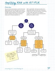
Cell Signaling Reverse Transcriptio
- Page 257 and 258:
Cell Signaling AMV Reverse Transcri
- Page 259 and 260:
Cell Signaling PCR Cloning pGEM ®
- Page 261 and 262:
Cell Signaling 15 Protein Expressio
- Page 263 and 264:
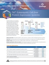
Cell Signaling TnT ® Quick Coupled
- Page 265 and 266:
Cell Signaling TnT ® T7 Quick for
- Page 267 and 268:
Cell Signaling Canine Pancreatic Mi
- Page 269 and 270:
Cell Signaling E. coli S30 Extract
- Page 271 and 272:
Cell Signaling Mass Spectrometry An
- Page 273 and 274:
Cell Signaling Endoproteinase Lys-C
- Page 275 and 276:
Cell Signaling Protein Labeling and
- Page 277 and 278:
Cell Signaling HaloTag ® Fusion (C
- Page 279 and 280:
Cell Signaling Inducible T7-Driven
- Page 281 and 282:
Cell Signaling ECL Western Blotting
- Page 283 and 284:
Cell Signaling 16 Protein Purificat
- Page 285 and 286:
Cell Signaling HaloTag ® Protein P
- Page 287 and 288:
HT Cell Signaling HaloTag ® Mammal
- Page 289 and 290:
Cell Signaling HaloLink Protein Arr
- Page 291 and 292:
Cell Signaling FastBreak Cell Lysis
- Page 293 and 294:
Cell Signaling MagneHis Protein Pur
- Page 295 and 296:
Cell Signaling Streptavidin Product
- Page 297 and 298:
Cell Signaling Protein Purification
- Page 299 and 300:
Cell Signaling 17 Reporter Assays a
- Page 301 and 302:
Cell Signaling NanoLuc Luciferase
- Page 303 and 304:
Cell Signaling Chroma-Glo Luciferas
- Page 305 and 306:
Cell Signaling ONE-Glo + Tox Lucife
- Page 307 and 308:
Cell Signaling Luciferase Assay Sys
- Page 309 and 310:
Cell Signaling ViviRen Live Cell Su
- Page 311 and 312:
Cell Signaling Reporter Vectors and
- Page 313 and 314:
Cell Signaling Promoterless Renilla
- Page 315 and 316:
Cell Signaling pmirGLO Dual-Lucifer
- Page 317 and 318:
Cell Signaling pGL2 Luciferase Repo
- Page 319 and 320:
Cell Signaling Reporter Vector Sequ
- Page 321 and 322:
Cell Signaling pCAT ® 3 Vectors Pr
- Page 323 and 324:
Cell Signaling ViviRen In Vivo Reni
- Page 325 and 326:
Cell Signaling 18 RNA Analysis 18 I
- Page 327 and 328:
Cell Signaling Riboprobe ® System
- Page 329 and 330:
Cell Signaling HeLaScribe ® Nuclea
- Page 331 and 332:
Cell Signaling RNA Interference Gen
- Page 333 and 334:
Cell Signaling 19 Stem Cell Researc
- Page 335 and 336:
Cell Signaling Cell ID System Fluor
- Page 337 and 338:
Cell Signaling StemElite Human Panc
- Page 339 and 340:
Cell Signaling 20 STR Analysis 20 C
- Page 341 and 342:
Cell Signaling PowerPlex ® 16 HS S
- Page 343 and 344:
Cell Signaling StemElite ID System
- Page 345 and 346:
Cell Signaling 21 Vectors 21 Bacter
- Page 347 and 348:
Cell Signaling Mammalian Expression
- Page 349 and 350:
Cell Signaling pCI-neo Mammalian Ex
- Page 351 and 352:
Cell Signaling Product Size Cat.# P
- Page 353 and 354:
Cell Signaling pSV-β-Galactosidase
- Page 355 and 356:
Cell Signaling Product Size Cat.# P
- Page 357 and 358:
22 Index 22 Index: A-Z 354 Index by
- Page 359 and 360:
Index: A-Z Click on name to go to p
- Page 361 and 362:
Index: A-Z Click on name to go to p
- Page 363 and 364:
Index: A-Z Click on name to go to p
- Page 365 and 366:
Index by Catalog Number Click on pa
- Page 367 and 368:
Index by Catalog Number Click on pa
- Page 369 and 370:
Index by Catalog Number Click on pa
- Page 371 and 372:
Index by Catalog Number Click on pa
- Page 373 and 374:
Index by Catalog Number Click on pa
- Page 375 and 376:
Index by Catalog Number Click on pa
- Page 377 and 378:
Index by Catalog Number Click on pa
- Page 379 and 380:
Index by Catalog Number Click on pa
- Page 381 and 382:
Index by Catalog Number Click on pa
- Page 383 and 384:
Index by Catalog Number Click on pa
- Page 385 and 386:
Index by Catalog Number Click on pa
- Page 387 and 388:
1S11 TH01 D3S1358 FGA TPOX D8S1179