

The genus Cladosporium and similar dematiaceous ... - CBS - KNAW
The genus Cladosporium and similar dematiaceous ... - CBS - KNAW
The genus Cladosporium and similar dematiaceous ... - CBS - KNAW

You also want an ePaper? Increase the reach of your titles
YUMPU automatically turns print PDFs into web optimized ePapers that Google loves.
<strong>Cladosporium</strong> herbarum species complex<br />
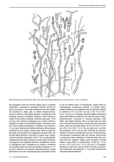
Fig. 13. <strong>Cladosporium</strong> herbaroides (CPC 12052). Macro- <strong>and</strong> micronematous conidiophores <strong>and</strong> conidia. Scale bar = 10 µm. K. Schubert del.<br />
like prolongation below the terminal swelling (due to sympodial<br />
proliferation), unbranched or sometimes branched, 30–230 µm<br />
long or even longer, 3–5 µm wide, swellings 5–8 µm wide, septate,<br />
not constricted at septa, pale to medium olivaceous-brown,<br />
smooth or almost so, walls slightly thickened. Conidiogenous cells<br />
integrated, terminal or intercalary, cylindrical, usually nodulose to<br />
nodose forming distinct swellings, sometimes geniculate, 15–55<br />
µm long, with numerous conidiogenous loci usually confined to<br />
swellings or situated on small lateral shoulders, sometimes on the<br />
top of short peg-like prolongations or denticles, loci protuberant,<br />
1–2 µm diam, thickened <strong>and</strong> darkened-refractive. Micronematous<br />
conidiophores much shorter, narrower, paler, neither nodulose nor<br />
geniculate, arising laterally from plagiotropous hyphae, often only<br />
as short lateral denticles or branchlets of hyphae, erect, straight,<br />
conical to cylindrical, unbranched, 3–65 × 2–3 µm, mostly aseptate,<br />
sometimes up to five septa, subhyaline, smooth, walls unthickened.<br />
Conidiogenous cells integrated, terminal or conidiophores reduced<br />
to conidiogenous cells, conidiogenous loci solitary or sometimes<br />
as sympodial clusters of pronounced denticles, protuberant, 1–1.5<br />
µm diam, thickened <strong>and</strong> somewhat darkened-refractive. Conidia<br />
polymorphous, two main morphological types recognisable, formed<br />
www.studiesinmycology.org<br />
by the two different types of conidiophores, conidia formed by<br />
macronematous conidiophores catenate, in branched chains,<br />
straight to slightly curved, subglobose, obovoid, limoniform, ellipsoid<br />
to cylindrical, 3–33 × (2–)3–6(–7) µm [av. ± SD, 14.5 (± 7.9) ×<br />
5.2 (± 1.2) µm], 0–2(–3)-septate, sometimes slightly constricted at<br />
septa, septa median or somewhat in the lower half, pale to medium<br />
olivaceous-brown, verruculose to verrucose (granulate under<br />
SEM), walls slightly thickened, with up to three rarely four distal<br />
scars, with age becoming medium or even dark brown (chocolate<br />
brown), wider <strong>and</strong> more thick-walled, 5.5–33 × (3.5–)5–9(–11) µm<br />
[av. ± SD, 14.4 (± 6.9) × 7.2 (± 1.9) µm], walls up to 1 µm thick,<br />
hila protuberant, 0.8–2(–2.5) µm diam, thickened <strong>and</strong> darkenedrefractive;<br />
microcyclic conidiogenesis occurring. Conidia formed by<br />
micronematous conidiophores paler <strong>and</strong> narrower, mostly formed<br />
in unbranched chains, sometimes in branched chains with up to<br />
three distal hila, straight to slightly curved, limoniform, narrowly<br />
fusiform, almost filiform to subcylindrical, 10–26(–35) × 2–3.5<br />
µm [av. ± SD, 15.6 (± 6.2) × 2.9 (± 0.5) µm], 0–1(–3)-septate,<br />
subhyaline to pale brown, almost smooth to minutely verruculose,<br />
walls unthickened, hila protuberant, 1–1.5 µm diam, thickened <strong>and</strong><br />
somewhat darkened-refractive.<br />
121


