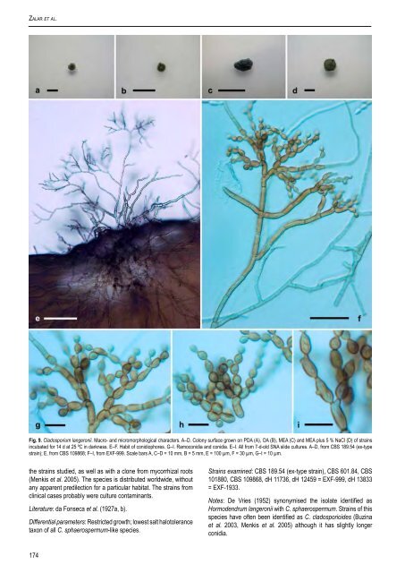
<strong>Cladosporium</strong> sphaerospermum species complex Mycelium partly submerged, partly superficial; hyphae without extracellular polysaccharide-like material. Conidiophores erect, arising laterally <strong>and</strong> terminally from straight hyphae, stipes of variable length, (5–)10–50(–300) × (2–)2.5–3(–5.5) μm, pale olivaceous-brown, smooth to minutely verruculose, thin-walled, 0– 3-septate, unbranched, with pronounced denticles. Conidial chains branching in all directions, terminal chains with up to 9 conidia. Conidiogenous cells undifferentiated. Ramoconidia rarely formed. Conidia verrucose, brown to dark brown, non-septate, usually subglobose to globose, less often short-ovoid, narrower at both ends, length : width ratio = 1.2–1.5; (2–)3–4(–6) × (2–)2.5–3(–5) μm [av. (± SD) 3.5 (± 0.7) × 2.7 (± 0.5)]; secondary ramoconidia cylindrical to almost spherical, 0–1-septate, (5–)7–12(–37.5) × (2–)2.5–3(–6.5) μm [av. (± SD) 10.3 (± 4.8) × 2.9 (± 0.6)], with up to 4 distal scars. Conidiogenous scars thickened <strong>and</strong> conspicuous, protuberant, 0.7–1.0(–1.5) μm diam. Cultural characteristics: Colonies on PDA reaching 27–43 mm diam, olive (2F5), slightly furrowed, often covered with grey secondary mycelium, except at the marginal area where only sporulating structures can be observed. Margin white <strong>and</strong> regular, with submerged hyphae. Reverse pale green to black. Colonies on OA reaching 29–40 mm diam, olive (2F6), flat, uniform, granular due to profuse sporulation <strong>and</strong> fasciculate bundles of conidiophores, without sterile mycelium. Reverse dark green to black. Colonies on MEA reaching 18–44 mm diam, highly variable in colour, but mainly olive (2E5), <strong>and</strong> from flat with regular margin to deeply furrowed with undulate margin. Colony centre wrinkled with crater-shaped appearance. Reverse pale to dark green. Colonies on MEA + 5 % NaCl reaching 24–48 mm diam, olive (3E8), furrowed, velvety, with more pale, undulate margins. Reverse dark green to black. Maximum tolerated salt concentration: Only 15 % of tested strains develop colonies at 20 % NaCl after 7 d, whereas after 14 d all cultures grow <strong>and</strong> sporulate. Cardinal temperatures: No growth at 4 °C, optimum 25 °C (18–44 mm diam), maximum 30 °C (6–23 mm diam). No growth at 37 °C. Specimen examined: Namibia, from hypersaline water of salterns, coll. Nina Gunde- Cimerman, 1 Sep. 2000, isol. P. Zalar, 1 Oct. 2000, <strong>CBS</strong> H-19734, holotype, culture ex-type EXF-572 = <strong>CBS</strong> 119416. Habitats <strong>and</strong> distribution: Hypersaline water in subtropical climates; indoor environments; Arctic ice; contaminant in lesions of humans <strong>and</strong> animals; plant phyllosphere; rock. Literature: Haubold et al. (1998), Meklin et al. (2004). Differential parameters: Verrucose conidia, short unbranched <strong>and</strong> non-septate conidiophores which arise laterally alongside erect hyphae. Strains examined: <strong>CBS</strong> 191.54, <strong>CBS</strong> 573.78, <strong>CBS</strong> 626.82, dH 12862, dH 12991, dH 13911, EXF-228, EXF-380, EXF-565, EXF- 567, EXF-571, EXF-572 (= <strong>CBS</strong> 119416; ex-type strain), EXF-646, EXF-698, EXF-703, EXF-944, EXF-972, EXF-977, EXF-1072, EXF-2372. Notes: <strong>Cladosporium</strong> halotolerans strongly resembles C. sphaerospermum. Several strains of this species such as dH 12862, dH 12941, <strong>CBS</strong> 191.54 <strong>and</strong> UAMH 7686 have been isolated sporadically from various indoor habitats in Europe, Brazil <strong>and</strong> the U.S.A. <strong>and</strong> repeatedly from bathrooms in Slovenia (Table 1). Probably sometimes as uncertain culture contaminations, it has been isolated from plants (GenBank accession no. L25433), www.studiesinmycology.org inner organs of a diseased frog (AY361982) <strong>and</strong> human brain (Kantarcioglu et al. 2002). <strong>The</strong> presence of C. halotolerans species in gypsum sediments entrapped in Arctic ice, the fact that it was repeatedly isolated from hypersaline water <strong>and</strong> possibly its presence in dolphin skin (see Discussion) suggest that it has a clear preference for (hyper)osmotic habitats. This is supported by its ability to grow at 20 % NaCl. <strong>The</strong> teleomorph of C. halotolerans is predicted to be a Davidiella species. Strain <strong>CBS</strong> 280.49 was isolated by J.A. von Arx from teleomorphic material of a fungus labelled as Mycosphaerella hyperici (Auersw.) Starbäck on Hypericum perforatum in Switzerl<strong>and</strong>. According to Aptroot (2006) this species may belong in Davidiella <strong>and</strong> produces a Septoria anamorph. In the original herbarium specimen, <strong>CBS</strong> H-4867, a Mycosphaerella teleomorph was present, but no sign of a <strong>Cladosporium</strong> anamorph. We assume that <strong>CBS</strong> 280.49 was a culture contaminant. <strong>Cladosporium</strong> langeronii (Fonseca, Leão & Nogueira) Vuill., Champ. Paras.: 78. 1931. Fig. 9. Basionym: Hormodendrum langeronii Fonseca, Leão & Nogueira, Sci. Med. 5: 563. 1927. ≡ <strong>Cladosporium</strong> langeronii (Fonseca, Leão & Nogueira) Cif., Manuale di Micologia Medica, ed. 2: 488 (1960), comb. superfl. Mycelium partly submerged, partly superficial; hyphae sometimes enveloped in polysaccharide-like material. Conidiophores erect or ascending, micronematous <strong>and</strong> macronematous, stipes of variable length, (20–)50–130(–200) × (3–)3.5–4.5(–6.5) μm, dark brown, rough- <strong>and</strong> thick-walled, regularly septate (cell length 9–22 μm), arising laterally <strong>and</strong> terminally from submerged or aerial hyphae, branched. Conidial chains dichotomously branched, up to 6 conidia in the unbranched parts. Conidiogenous cells undifferentiated, sometimes seceding <strong>and</strong> forming ramoconidia. Ramoconidia cylindrical, 0–1 septate, (10–)11–22(–42) × (3–)3.5–4.5(–5) µm, base broadly truncate, 2–3.5 µm wide, slightly thickened <strong>and</strong> somewhat darkened. Conidia irregularly verruculose to sometimes loosely verrucose, dark brown, non-septate, usually ovoid, length : width ratio = 1.3–1.5; conidial size (3–)4–5.5(–8) × (2–)3–4(–5) μm [av. (± SD) 4.8 (± 1.0) × 3.5 (± 0.6)]; secondary ramoconidia cylindrical to almost spherical, mostly 0–1(–2)-septate, (5.5–)7.5– 12.5(–35.5) × (2.5–)3–4.5(–5.5) μm [av. (± SD) 10.7 (± 4.7) × 3.6 (± 0.8)], with 2, rarely 3 distal scars. Conidiogenous scars thickened <strong>and</strong> conspicuous, protuberant, 0.9–1.5(–2.3) μm diam. Cultural characteristics: Colonies on PDA, OA <strong>and</strong> MEA with restricted growth, attaining 2.5–4.5, 1.5–7.0 <strong>and</strong> 1.0–5.5 mm diam, respectively. Colonies flat or heaped (up to 3 mm), dark green (30F4), with black reverse <strong>and</strong> slightly undulate margin with immersed mycelium. Sporulating on all media. On MEA + 5 % NaCl growth is faster, colonies attaining 8.5–12.0 mm diam, sporulating <strong>and</strong> growing deeply into the agar. Maximum tolerated salt concentration: All strains develop colonies at 17 % NaCl after 14 d. Cardinal temperatures: No growth at 4 °C, optimum / maximum 25 °C (1.0–5.5 mm diam), no growth at 30 °C. Specimen examined: Brazil, from man ulcero-nodular mycosis of h<strong>and</strong> <strong>and</strong> arm, 1927, coll. <strong>and</strong> isol. da Fonseca, <strong>CBS</strong> H-19737, holotype, culture ex-type <strong>CBS</strong> 189.54. Habitats <strong>and</strong> distribution: Polar ice <strong>and</strong> biomats; conifer wood <strong>and</strong> window frame in Europe; humans; strains originating from nasal mucus (Buzina et al. 2003) have 100 % sequence homology with 173
Zalar et al. Fig. 9. <strong>Cladosporium</strong> langeronii. Macro- <strong>and</strong> micromorphological characters. A–D. Colony surface grown on PDA (A), OA (B), MEA (C) <strong>and</strong> MEA plus 5 % NaCl (D) of strains incubated for 14 d at 25 ºC in darkness. E–F. Habit of conidiophores. G–I. Ramoconidia <strong>and</strong> conidia. E–I. All from 7-d-old SNA slide cultures. A–D, from <strong>CBS</strong> 189.54 (ex-type strain); E, from <strong>CBS</strong> 109868; F–I, from EXF-999. Scale bars A, C–D = 10 mm, B = 5 mm, E = 100 µm, F = 30 µm, G–I = 10 µm. the strains studied, as well as with a clone from mycorrhizal roots (Menkis et al. 2005). <strong>The</strong> species is distributed worldwide, without any apparent predilection for a particular habitat. <strong>The</strong> strains from clinical cases probably were culture contaminants. Literature: da Fonseca et al. (1927a, b). Differential parameters: Restricted growth; lowest salt halotolerance taxon of all C. sphaerospermum-like species. Strains examined: <strong>CBS</strong> 189.54 (ex-type strain), <strong>CBS</strong> 601.84, <strong>CBS</strong> 101880, <strong>CBS</strong> 109868, dH 11736, dH 12459 = EXF-999, dH 13833 = EXF-1933. Notes: De Vries (1952) synonymised the isolate identified as Hormodendrum langeronii with C. sphaerospermum. Strains of this species have often been identified as C. cladosporioides (Buzina et al. 2003, Menkis et al. 2005) although it has slightly longer conidia. 174
- Page 1:
Studies in Mycology 58 (2007) The g
- Page 4 and 5:
Studies in Mycology The Studies in
- Page 7 and 8:
CONTENTS P.W. Crous, U. Braun and J
- Page 9 and 10:
lectotype for the genus by Clements
- Page 11:
Schubert K (2005a). Morphotaxonomic
- Page 14 and 15:
Crous et al. Table 1. Isolates for
- Page 16 and 17:
Crous et al. Table 1. (Continued).
- Page 18 and 19:
Crous et al. 100 10 changes 65 100
- Page 20 and 21:
Crous et al. Treatment of phylogene
- Page 22 and 23:
Crous et al. Teratosphaeria bellula
- Page 24 and 25:
Crous et al. 6. Conidiophores short
- Page 26 and 27:
Crous et al. Habit plant pathogenic
- Page 28 and 29:
Crous et al. Fig. 7. Catenulostroma
- Page 30 and 31:
Crous et al. system, consisting of
- Page 32 and 33:
Crous et al. Fig. 9. Penidiella col
- Page 34 and 35:
Crous et al. Fig. 12. Penidiella ri
- Page 36 and 37:
Crous et al. conidiophores, about 1
- Page 38 and 39:
Crous et al. have conidiomata rangi
- Page 40 and 41:
Crous et al. Schizothyriaceae clade
- Page 42 and 43:
Crous et al. Fig. 21. Stigmidium sc
- Page 44 and 45:
Crous et al. Gams W, Verkley GJM, C
- Page 46 and 47:
Crous et al. Table 1. Isolates for
- Page 48 and 49:
Crous et al. 100 61 100 52 100 10 c
- Page 50 and 51:
Crous et al. Fig. 3. Rachicladospor
- Page 52 and 53:
Crous et al. Fig. 4. Toxicocladospo
- Page 54 and 55:
Crous et al. Fig. 5. Verrucocladosp
- Page 56 and 57:
Crous et al. Fig. 7. Stenella aragu
- Page 58 and 59:
Crous et al. Helotiales, incertae s
- Page 60 and 61:
Crous et al. Fig. 10. Ochrocladospo
- Page 62 and 63:
Crous et al. Fig. 12. Rhizocladospo
- Page 64 and 65:
Crous et al. 7. Conidiophores unbra
- Page 66 and 67:
Crous et al. 33. Terminal conidioge
- Page 68 and 69:
Crous et al. Crous PW, Kang JC, Bra
- Page 70 and 71:
Arzanlou et al. To date 26 species
- Page 72 and 73:
Arzanlou et al. Table 1. (Continued
- Page 74 and 75:
Arzanlou et al. 10 changes Athelia
- Page 76 and 77:
Arzanlou et al. Athelia epiphylla A
- Page 78 and 79:
Arzanlou et al. Fig. 3. Periconiell
- Page 80 and 81:
Arzanlou et al. Cultural characteri
- Page 82 and 83:
Arzanlou et al. Fig. 10. A. Ramichl
- Page 84 and 85:
Arzanlou et al. Ramichloridium musa
- Page 86 and 87:
Arzanlou et al. Cultural characteri
- Page 88 and 89:
Arzanlou et al. Fig. 20. Rhinocladi
- Page 90 and 91:
Arzanlou et al. Fig. 22. Rhinocladi
- Page 92 and 93:
Arzanlou et al. Rhinocladiella mack
- Page 94 and 95:
Arzanlou et al. Fig. 26. Veronaea c
- Page 96 and 97:
Arzanlou et al. Cultural characteri
- Page 98 and 99:
Arzanlou et al. Fig. 30. Myrmecridi
- Page 100 and 101:
Arzanlou et al. Fig. 32. Radulidium
- Page 102 and 103:
Arzanlou et al. Fig. 34. Rhodoveron
- Page 104 and 105:
Arzanlou et al. even further, thoug
- Page 106 and 107:
available online at www.studiesinmy
- Page 108 and 109:
Dichocladosporium gen. nov. 10 chan
- Page 110 and 111:
Dichocladosporium gen. nov. Fig. 3.
- Page 112 and 113:
Dichocladosporium gen. nov. to intr
- Page 114 and 115:
Dichocladosporium gen. nov. Czechos
- Page 116 and 117:
available online at www.studiesinmy
- Page 118 and 119:
Cladosporium herbarum species compl
- Page 120 and 121:
Cladosporium herbarum species compl
- Page 122 and 123:
Cladosporium herbarum species compl
- Page 124 and 125:
Cladosporium herbarum species compl
- Page 126 and 127:
Cladosporium herbarum species compl
- Page 128 and 129:
Cladosporium herbarum species compl
- Page 130 and 131:
Cladosporium herbarum species compl
- Page 132 and 133:
Cladosporium herbarum species compl
- Page 134 and 135: Cladosporium herbarum species compl
- Page 136 and 137: Cladosporium herbarum species compl
- Page 138 and 139: Cladosporium herbarum species compl
- Page 140 and 141: Cladosporium herbarum species compl
- Page 142 and 143: Cladosporium herbarum species compl
- Page 144 and 145: Cladosporium herbarum species compl
- Page 146 and 147: Cladosporium herbarum species compl
- Page 148 and 149: Cladosporium herbarum species compl
- Page 150 and 151: Cladosporium herbarum species compl
- Page 152 and 153: Cladosporium herbarum species compl
- Page 154 and 155: Cladosporium herbarum species compl
- Page 156 and 157: Cladosporium herbarum species compl
- Page 158 and 159: Cladosporium herbarum species compl
- Page 160 and 161: Cladosporium herbarum species compl
- Page 162 and 163: Cladosporium herbarum species compl
- Page 164 and 165: Cladosporium herbarum species compl
- Page 166 and 167: Cladosporium herbarum species compl
- Page 168 and 169: available online at www.studiesinmy
- Page 170 and 171: Cladosporium sphaerospermum species
- Page 172 and 173: Cladosporium sphaerospermum species
- Page 174 and 175: Cladosporium sphaerospermum species
- Page 176 and 177: Cladosporium sphaerospermum species
- Page 178 and 179: Cladosporium sphaerospermum species
- Page 180 and 181: Cladosporium sphaerospermum species
- Page 182 and 183: Cladosporium sphaerospermum species
- Page 186 and 187: Cladosporium sphaerospermum species
- Page 188 and 189: Cladosporium sphaerospermum species
- Page 190 and 191: Cladosporium sphaerospermum species
- Page 192 and 193: Cladosporium sphaerospermum species
- Page 194 and 195: Cladosporium sphaerospermum species
- Page 196 and 197: Crous et al. The aim of the present
- Page 198 and 199: Crous et al. 10 changes Athelia epi
- Page 200 and 201: Crous et al. Table 1. Isolates used
- Page 202 and 203: Crous et al. Table 1. (Continued).
- Page 204 and 205: Crous et al. 0.1 expected changes p
- Page 206 and 207: Crous et al. Fig. 6. Cladophialopho
- Page 208 and 209: Crous et al. Fig. 11. Cladophialoph
- Page 210 and 211: Crous et al. Fig. 13. Cladophialoph
- Page 212 and 213: Crous et al. Fig. 15. Exophiala sp.
- Page 214 and 215: Crous et al. Fig. 18. Cylindrosympo
- Page 216 and 217: Crous et al. Fig. 19. Fusicladium a
- Page 218 and 219: Crous et al. Fig. 22. Fusicladium f
- Page 220 and 221: Crous et al. Fig. 24. Fusicladium p
- Page 222 and 223: Crous et al. Fusicladium rhodense C
- Page 224 and 225: Crous et al. Excluded taxa Polyscyt
- Page 226 and 227: Crous et al. the conidial tips are
- Page 228 and 229: available online at www.studiesinmy
- Page 230 and 231: Cladophialophora carrionii complex
- Page 232 and 233: Cladophialophora carrionii complex
- Page 234 and 235:
Cladophialophora carrionii complex
- Page 236 and 237:
Cladophialophora carrionii complex
- Page 238 and 239:
Cladophialophora carrionii complex
- Page 240 and 241:
Cladophialophora carrionii complex
- Page 242 and 243:
Cladophialophora carrionii complex
- Page 244 and 245:
available online at www.studiesinmy
- Page 246 and 247:
Hormoconis resinae and morphologica
- Page 248 and 249:
Hormoconis resinae and morphologica
- Page 250 and 251:
Hormoconis resinae and morphologica
- Page 252 and 253:
Fig. 6 Hormoconis resinae and morph
- Page 254 and 255:
Hormoconis resinae and morphologica
- Page 256 and 257:
Cladophialophora, 52 k , 54 k -55,
- Page 258 and 259:
Fusicladium phillyreae, 189 c , 191
- Page 260 and 261:
Ramichloridium epichloës, 60, 89 P
- Page 262:
Trimmatostroma salicis, 3 t , 5, 6