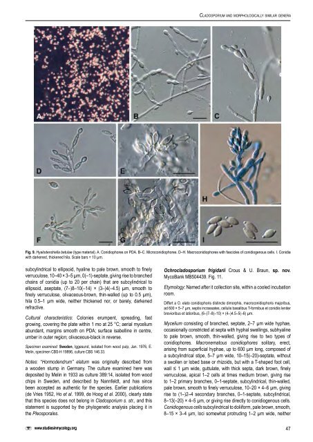
Crous et al. Helotiales, incertae sedis Hyalodendriella Crous, gen. nov. MycoBank MB504435. Etymology: Morphologically <strong>similar</strong> to Hyalodendron Diddens. Differt a Hyalodendro et Retroconi conidiophoris dimorphis, cicatricibus incrassatis et conidiis ultimo brunneis. Morphologically <strong>similar</strong> to Hyalodendron <strong>and</strong> Retroconis, but distinct in that it has dimorphic conidiophores, conidia that turn brown with age, <strong>and</strong> have thickened scars. Microconidiophores forming as lateral branches on hyphae, subcylindrical, subhyaline to pale brown, smooth, septate, with terminal conidiogenous cells. Macroconidiophores septate, subcylindrical, straight to curved, subhyaline to pale brown, smooth, with an apical rachis that is pale brown, smooth, subcylindrical, with numerous, aggregated loci. Conidia limoniform to ellipsoid, aseptate, smooth, pale brown, in short chains, tapering towards ends that are prominently apiculate, prominently thickened <strong>and</strong> darkened, but not refractive. Type species: Hyalodendriella betulae Crous, sp. nov. Hyalodendriella betulae Crous sp. nov. MycoBank MB504436. Fig. 9. Mycelium ex hyphis ramosis, septatis, 1.5–2 µm latis, levibus, hyalinis vel pallide brunnei compositum. Conidiophora dimorphosa: (A) Conidiophora ex hyphis lateraliter oriunda, subcylindrical, subhyalina vel pallide brunnea, levia, 1–6- septata, ad 40 µm longa et 2–3 µm lata. Cellulae conidiogenae terminales, 5–15 × 2–3 µm, loco conidiogeno singulare et terminale, cellula ellipsoidea (conidio ?), persistente, interdum cellulis catenulatis (ad 6), pallide brunneo, apice subacute rotundato, basi truncata, 5–7 × 3–4 µm. (B) Conidiophoris 10–20 × 2–3 µm, 1–2-septatis, subcylindraceis, rectis vel curvatis, subhyalinis vel pallide brunneis, levibus. Cellulae conidiogenae pallide brunneae, leviae, subcylindraceae, locis numerosis, aggregatis, inconspicuis vel subdenticulatis, leviter protuberantes, 0.5 µm diam, incrassatis et fuscatis. Conidia catenulata (2–3), (4–)5–6(–7) × 2.5–3 µm, limoniformes vel ellipsoidea, non septata, levia, pallide brunnea, utrinque attenuata, apiculata, 0.5–1 × 0.5 µm, incrassata et fuscata, non refractiva. Mycelium consisting of branched, septate, 1.5–2 µm wide hyphae, smooth, hyaline to pale brown. Conidiophores dimorphic. Type A: Conidiophores forming as lateral branches on hyphae, subcylindrical, subhyaline to pale brown, smooth, 1–6-septate, up to 40 µm long, <strong>and</strong> 2–3 µm wide. Conidiogenous cells terminal, 5– 15 × 2–3 µm, with a single, apical locus, giving rise to an ellipsoidal cell (conidium?) which mostly remains attached, pale brown, with a subacutely rounded apex <strong>and</strong> truncate base, 5–7 × 3–4 µm, at times forming chains of up to 6 such cells. Type B: Conidiophores 10–20 × 2–3 µm, 1–2-septate, subcylindrical, straight to curved, subhyaline to pale brown, smooth. Conidiogenous cells pale brown, smooth, subcylindrical with numerous, aggregated loci, inconspicuous to subdenticulate <strong>and</strong> somewhat protruding, 0.5 µm wide, somewhat thickened <strong>and</strong> darkened. Conidia in chains of 2–3, limoniform to ellipsoid, widest in the middle, aseptate, smooth, pale brown, tapering towards ends that are prominently apiculate, 0.5–1 µm long, 0.5 µm wide, prominently thickened <strong>and</strong> darkened, but not refractive. Cultural characteristics: Colonies on PDA slimy, spreading, somewhat erumpent in the centre, with even, catenulate margins, lacking aerial mycelium; surface fuscous-black to olivaceous-black, with patches of cream; reverse fuscous-black with patches of cream. Colonies reaching 25 mm diam on PDA after 1 mo at 25 °C in the dark; colonies fertile with profuse sporulation on SNA. Specimen examined: Netherl<strong>and</strong>s, Oostelijk Flevol<strong>and</strong>, Jagersveld, isolated from Alnus glutinosa (Betulaceae), May 1982, W. Gams, holotype <strong>CBS</strong>-H 19895, culture ex-type <strong>CBS</strong> 261.82. Notes: Morphologically Hyalodendriella resembles the genera Hyalodendron <strong>and</strong> Retroconis de Hoog & Bat. Vegte (de Hoog & Batenburg van der Vegte 1989). It is distinct, however, in its pigmentation, dimorphic conidiophores <strong>and</strong> conidia. Furthermore, a strain of Retroconis fusiformis (S.M. Reddy & Bilgrami) de Hoog & Bat. Vegte (<strong>CBS</strong> 330.81) clusters apart from Hyalodendriella, namely in the Chaetomiaceae, Sordariales. Pleosporales, incertae sedis Ochrocladosporium Crous & U. Braun, gen. nov. MycoBank MB504437. Etymology: Named after its pale brown, cladosporium-like conidia. Differt a Cladosporio et generis cladosporioidibus diversis conidiophoris cum cellulis basalibus T-formibus et/vel cicatricibus non incrassatis, non vel leviter fuscatisrefractivis. Mycelium consisting of branched, septate hyphae, subhyaline to pale brown, smooth, giving rise to two types of conidiophores. Macronematous conidiophores solitary, erect, arising from superficial hyphae, composed of a subcylindrical stipe, without a swollen or lobed base or rhizoids, with or without a T-shaped foot cell, pale to dark brown; apical conidiogenous apparatus with or without additional branches, branched part, if present, with short branchlets composed of conidiogenous cells <strong>and</strong> ramoconidia, continuous to septate, wall thin or slightly thicked, pale brown. Conidiogenous cells integrated, terminal or intercalary, subcylindrical to doliiform, pale brown, thin-walled, smooth; unilocal or multilocal, determinate to sympodial, loci conically truncate, subdenticulate, neither thickened, nor darkened-refractive or only slightly darkened-refractive. Micronematous conidiophores integrated in hyphae, reduced to a lateral peg-like locus or erect, frequently reduced to conidiogenous cells, pale brown, smooth, subcylindrical. Conidia occurring in branched chains, fusiform, ellipsoid-ovoid to subcylindrical, 0(–1)- septate, ramoconidia present, pale brown, thin-walled, smooth to finely verruculose, ends attenuated, hila obconically truncate to almost pointed, neither thickened nor darkened-refractive. Type species: Ochrocladosporium elatum (Harz) Crous & U. Braun, comb. nov. Ochrocladosporium elatum (Harz) Crous & U. Braun, comb. nov. MycoBank MB504438. Fig. 10. Basionym: Hormodendrum elatum Harz, Bull. Soc. Imp. Naturalistes Moscou 44: 140. 1871. ≡ <strong>Cladosporium</strong> elatum (Harz) Nannf., in Melin & Nannfeldt, Svenska Skogsvardsfoereren Tidskr. 32: 397. 1934. ≡ Cadophora elatum (Harz) Nannf., in Melin & Nannfeldt, Svenska Skogsvardsfoereren Tidskr. 32: 422. 1934. Mycelium consisting of branched, septate, smooth, hyaline, 2–4 µm wide, thin-walled, hyphae, becoming darker brown in places, giving rise to erect conidiophores. Conidiophores either reduced to conidiogenous cells, or well-differentiated, terminal <strong>and</strong> lateral on hyphae, erect, highly variable, arising from superficial <strong>and</strong> submerged hyphae, reduced to subdenticulate loci, 1–1.5 µm wide, or well-differentiated, up to 60 µm long, 1–3-septate, 3–4 µm wide, hyaline to medium brown, smooth, thin-walled (≤ 1 µm). Conidiogenous cells integrated as lateral peg-like loci on hyphal cells, or erect, subcylindrical, up to 25 µm long, 2.5–4 µm wide, with 1–3 terminal loci, occasionally also lateral, 1–1.5 µm wide, not thickened <strong>and</strong> darkened, but frequently somewhat refractive (mounted in Shear’s solution, not lactic acid). Ramoconidia 46
<strong>Cladosporium</strong> <strong>and</strong> morphologically <strong>similar</strong> genera Fig. 9. Hyalodendriella betulae (type material). A. Conidiophores on PDA. B–C. Microconidiophores. D–H. Macroconidiophores with fascicles of conidiogenous cells. I. Conidia with darkened, thickened hila. Scale bars = 10 µm. subcylindrical to ellipsoid, hyaline to pale brown, smooth to finely verruculose, 10–40 × 3–5 µm, 0(–1)-septate, giving rise to branched chains of conidia (up to 20 per chain) that are subcylindrical to ellipsoid, aseptate, (7–)8–10(–14) × (3–)4(–4.5) µm, smooth to finely verruculose, olivaceous-brown, thin-walled (up to 0.5 µm), hila 0.5–1 µm wide, neither thickened nor, or barely, darkened refractive. Cultural characteristics: Colonies erumpent, spreading, fast growing, covering the plate within 1 mo at 25 °C; aerial mycelium abundant, margins smooth on PDA; surface isabelline in centre, umber in outer region; olivaceous-black in reverse. Specimen examined: Sweden, Iggesund, isolated from wood pulp, Jan. 1976, E. Melin, specimen <strong>CBS</strong>-H 19896, culture <strong>CBS</strong> 146.33. Notes: “Hormodendrum” elatum was originally described from a wooden stump in Germany. <strong>The</strong> culture examined here was deposited by Melin in 1933 as culture 389:14, isolated from wood chips in Sweden, <strong>and</strong> described by Nannfeldt, <strong>and</strong> has since been accepted as authentic for the species. Earlier publications (de Vries 1952, Ho et al. 1999, de Hoog et al. 2000), clearly state that this species does not belong in <strong>Cladosporium</strong> s. str., <strong>and</strong> this statement is supported by the phylogenetic analysis placing it in the Pleosporales. www.studiesinmycology.org Ochrocladosporium frigidarii Crous & U. Braun, sp. nov. MycoBank MB504439. Fig. 11. Etymology: Named after it collection site, within a cooled incubation room. Differt a O. elato conidiophoris distincte dimorphis, macroconidiophoris majoribus, ad 600 × 5–7 µm, septis incrassates, cellulis basalibus T-formibus et conidiis leniter brevioribus et latioribus, (6–)7–8(–10) × (4–)4.5–5(–6) µm. Mycelium consisting of branched, septate, 2–7 µm wide hyphae, occasionally constricted at septa with hyphal swellings, subhyaline to pale brown, smooth, thin-walled, giving rise to two types of conidiophores. Macronematous conidiophores solitary, erect, arising from superficial hyphae, up to 600 µm long, composed of a subcylindrical stipe, 5–7 µm wide, 10–15(–20)-septate, without a swollen or lobed base or rhizoids, but with a T-shaped foot cell, wall ≤ 1 µm wide, guttulate, with thick septa, dark brown, finely verruculose, apical 1–2 cells at times medium brown, giving rise to 1–2 primary branches, 0–1-septate, subcylindrical, thin-walled, pale brown, smooth to finely verruculose, 10–20 × 4–6 µm, giving rise to (1–)2–4 secondary branches, 0–1-septate, subcylindrical, 8–13(–20) × 4–5 µm, or giving rise directly to conidiogenous cells. Conidiogenous cells subcylindrical to doliiform, pale brown, smooth, 8–15 × 3–4 µm, loci somewhat protruding 1–2 µm wide, neither 47
- Page 1:
Studies in Mycology 58 (2007) The g
- Page 4 and 5:
Studies in Mycology The Studies in
- Page 7 and 8: CONTENTS P.W. Crous, U. Braun and J
- Page 9 and 10: lectotype for the genus by Clements
- Page 11: Schubert K (2005a). Morphotaxonomic
- Page 14 and 15: Crous et al. Table 1. Isolates for
- Page 16 and 17: Crous et al. Table 1. (Continued).
- Page 18 and 19: Crous et al. 100 10 changes 65 100
- Page 20 and 21: Crous et al. Treatment of phylogene
- Page 22 and 23: Crous et al. Teratosphaeria bellula
- Page 24 and 25: Crous et al. 6. Conidiophores short
- Page 26 and 27: Crous et al. Habit plant pathogenic
- Page 28 and 29: Crous et al. Fig. 7. Catenulostroma
- Page 30 and 31: Crous et al. system, consisting of
- Page 32 and 33: Crous et al. Fig. 9. Penidiella col
- Page 34 and 35: Crous et al. Fig. 12. Penidiella ri
- Page 36 and 37: Crous et al. conidiophores, about 1
- Page 38 and 39: Crous et al. have conidiomata rangi
- Page 40 and 41: Crous et al. Schizothyriaceae clade
- Page 42 and 43: Crous et al. Fig. 21. Stigmidium sc
- Page 44 and 45: Crous et al. Gams W, Verkley GJM, C
- Page 46 and 47: Crous et al. Table 1. Isolates for
- Page 48 and 49: Crous et al. 100 61 100 52 100 10 c
- Page 50 and 51: Crous et al. Fig. 3. Rachicladospor
- Page 52 and 53: Crous et al. Fig. 4. Toxicocladospo
- Page 54 and 55: Crous et al. Fig. 5. Verrucocladosp
- Page 56 and 57: Crous et al. Fig. 7. Stenella aragu
- Page 60 and 61: Crous et al. Fig. 10. Ochrocladospo
- Page 62 and 63: Crous et al. Fig. 12. Rhizocladospo
- Page 64 and 65: Crous et al. 7. Conidiophores unbra
- Page 66 and 67: Crous et al. 33. Terminal conidioge
- Page 68 and 69: Crous et al. Crous PW, Kang JC, Bra
- Page 70 and 71: Arzanlou et al. To date 26 species
- Page 72 and 73: Arzanlou et al. Table 1. (Continued
- Page 74 and 75: Arzanlou et al. 10 changes Athelia
- Page 76 and 77: Arzanlou et al. Athelia epiphylla A
- Page 78 and 79: Arzanlou et al. Fig. 3. Periconiell
- Page 80 and 81: Arzanlou et al. Cultural characteri
- Page 82 and 83: Arzanlou et al. Fig. 10. A. Ramichl
- Page 84 and 85: Arzanlou et al. Ramichloridium musa
- Page 86 and 87: Arzanlou et al. Cultural characteri
- Page 88 and 89: Arzanlou et al. Fig. 20. Rhinocladi
- Page 90 and 91: Arzanlou et al. Fig. 22. Rhinocladi
- Page 92 and 93: Arzanlou et al. Rhinocladiella mack
- Page 94 and 95: Arzanlou et al. Fig. 26. Veronaea c
- Page 96 and 97: Arzanlou et al. Cultural characteri
- Page 98 and 99: Arzanlou et al. Fig. 30. Myrmecridi
- Page 100 and 101: Arzanlou et al. Fig. 32. Radulidium
- Page 102 and 103: Arzanlou et al. Fig. 34. Rhodoveron
- Page 104 and 105: Arzanlou et al. even further, thoug
- Page 106 and 107: available online at www.studiesinmy
- Page 108 and 109:
Dichocladosporium gen. nov. 10 chan
- Page 110 and 111:
Dichocladosporium gen. nov. Fig. 3.
- Page 112 and 113:
Dichocladosporium gen. nov. to intr
- Page 114 and 115:
Dichocladosporium gen. nov. Czechos
- Page 116 and 117:
available online at www.studiesinmy
- Page 118 and 119:
Cladosporium herbarum species compl
- Page 120 and 121:
Cladosporium herbarum species compl
- Page 122 and 123:
Cladosporium herbarum species compl
- Page 124 and 125:
Cladosporium herbarum species compl
- Page 126 and 127:
Cladosporium herbarum species compl
- Page 128 and 129:
Cladosporium herbarum species compl
- Page 130 and 131:
Cladosporium herbarum species compl
- Page 132 and 133:
Cladosporium herbarum species compl
- Page 134 and 135:
Cladosporium herbarum species compl
- Page 136 and 137:
Cladosporium herbarum species compl
- Page 138 and 139:
Cladosporium herbarum species compl
- Page 140 and 141:
Cladosporium herbarum species compl
- Page 142 and 143:
Cladosporium herbarum species compl
- Page 144 and 145:
Cladosporium herbarum species compl
- Page 146 and 147:
Cladosporium herbarum species compl
- Page 148 and 149:
Cladosporium herbarum species compl
- Page 150 and 151:
Cladosporium herbarum species compl
- Page 152 and 153:
Cladosporium herbarum species compl
- Page 154 and 155:
Cladosporium herbarum species compl
- Page 156 and 157:
Cladosporium herbarum species compl
- Page 158 and 159:
Cladosporium herbarum species compl
- Page 160 and 161:
Cladosporium herbarum species compl
- Page 162 and 163:
Cladosporium herbarum species compl
- Page 164 and 165:
Cladosporium herbarum species compl
- Page 166 and 167:
Cladosporium herbarum species compl
- Page 168 and 169:
available online at www.studiesinmy
- Page 170 and 171:
Cladosporium sphaerospermum species
- Page 172 and 173:
Cladosporium sphaerospermum species
- Page 174 and 175:
Cladosporium sphaerospermum species
- Page 176 and 177:
Cladosporium sphaerospermum species
- Page 178 and 179:
Cladosporium sphaerospermum species
- Page 180 and 181:
Cladosporium sphaerospermum species
- Page 182 and 183:
Cladosporium sphaerospermum species
- Page 184 and 185:
Cladosporium sphaerospermum species
- Page 186 and 187:
Cladosporium sphaerospermum species
- Page 188 and 189:
Cladosporium sphaerospermum species
- Page 190 and 191:
Cladosporium sphaerospermum species
- Page 192 and 193:
Cladosporium sphaerospermum species
- Page 194 and 195:
Cladosporium sphaerospermum species
- Page 196 and 197:
Crous et al. The aim of the present
- Page 198 and 199:
Crous et al. 10 changes Athelia epi
- Page 200 and 201:
Crous et al. Table 1. Isolates used
- Page 202 and 203:
Crous et al. Table 1. (Continued).
- Page 204 and 205:
Crous et al. 0.1 expected changes p
- Page 206 and 207:
Crous et al. Fig. 6. Cladophialopho
- Page 208 and 209:
Crous et al. Fig. 11. Cladophialoph
- Page 210 and 211:
Crous et al. Fig. 13. Cladophialoph
- Page 212 and 213:
Crous et al. Fig. 15. Exophiala sp.
- Page 214 and 215:
Crous et al. Fig. 18. Cylindrosympo
- Page 216 and 217:
Crous et al. Fig. 19. Fusicladium a
- Page 218 and 219:
Crous et al. Fig. 22. Fusicladium f
- Page 220 and 221:
Crous et al. Fig. 24. Fusicladium p
- Page 222 and 223:
Crous et al. Fusicladium rhodense C
- Page 224 and 225:
Crous et al. Excluded taxa Polyscyt
- Page 226 and 227:
Crous et al. the conidial tips are
- Page 228 and 229:
available online at www.studiesinmy
- Page 230 and 231:
Cladophialophora carrionii complex
- Page 232 and 233:
Cladophialophora carrionii complex
- Page 234 and 235:
Cladophialophora carrionii complex
- Page 236 and 237:
Cladophialophora carrionii complex
- Page 238 and 239:
Cladophialophora carrionii complex
- Page 240 and 241:
Cladophialophora carrionii complex
- Page 242 and 243:
Cladophialophora carrionii complex
- Page 244 and 245:
available online at www.studiesinmy
- Page 246 and 247:
Hormoconis resinae and morphologica
- Page 248 and 249:
Hormoconis resinae and morphologica
- Page 250 and 251:
Hormoconis resinae and morphologica
- Page 252 and 253:
Fig. 6 Hormoconis resinae and morph
- Page 254 and 255:
Hormoconis resinae and morphologica
- Page 256 and 257:
Cladophialophora, 52 k , 54 k -55,
- Page 258 and 259:
Fusicladium phillyreae, 189 c , 191
- Page 260 and 261:
Ramichloridium epichloës, 60, 89 P
- Page 262:
Trimmatostroma salicis, 3 t , 5, 6