

The genus Cladosporium and similar dematiaceous ... - CBS - KNAW
The genus Cladosporium and similar dematiaceous ... - CBS - KNAW
The genus Cladosporium and similar dematiaceous ... - CBS - KNAW

You also want an ePaper? Increase the reach of your titles
YUMPU automatically turns print PDFs into web optimized ePapers that Google loves.
Ramichloridium <strong>and</strong> allied genera<br />
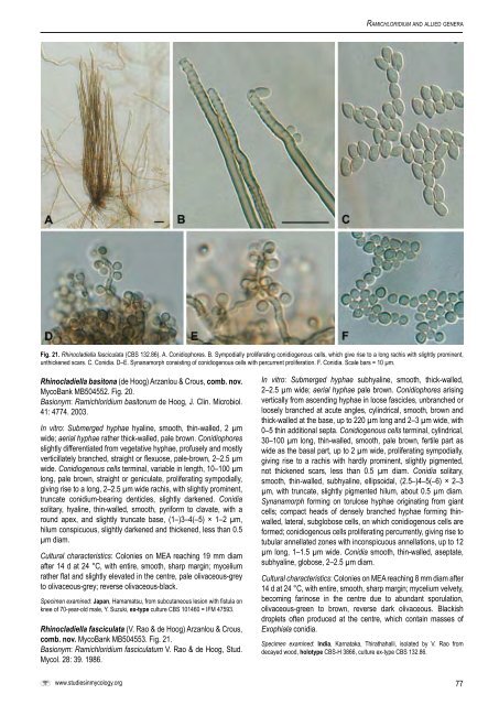
Fig. 21. Rhinocladiella fasciculata (<strong>CBS</strong> 132.86). A. Conidiophores. B. Sympodially proliferating conidiogenous cells, which give rise to a long rachis with slightly prominent,<br />
unthickened scars. C. Conidia. D–E. Synanamorph consisting of conidiogenous cells with percurrent proliferation. F. Conidia. Scale bars = 10 µm.<br />
Rhinocladiella basitona (de Hoog) Arzanlou & Crous, comb. nov.<br />
MycoBank MB504552. Fig. 20.<br />
Basionym: Ramichloridium basitonum de Hoog, J. Clin. Microbiol.<br />
41: 4774. 2003.<br />
In vitro: Submerged hyphae hyaline, smooth, thin-walled, 2 µm<br />
wide; aerial hyphae rather thick-walled, pale brown. Conidiophores<br />
slightly differentiated from vegetative hyphae, profusely <strong>and</strong> mostly<br />
verticillately branched, straight or flexuose, pale-brown, 2–2.5 µm<br />
wide. Conidiogenous cells terminal, variable in length, 10–100 µm<br />
long, pale brown, straight or geniculate, proliferating sympodially,<br />
giving rise to a long, 2–2.5 µm wide rachis, with slightly prominent,<br />
truncate conidium-bearing denticles, slightly darkened. Conidia<br />
solitary, hyaline, thin-walled, smooth, pyriform to clavate, with a<br />
round apex, <strong>and</strong> slightly truncate base, (1–)3–4(–5) × 1–2 µm,<br />
hilum conspicuous, slightly darkened <strong>and</strong> thickened, less than 0.5<br />
µm diam.<br />
Cultural characteristics: Colonies on MEA reaching 19 mm diam<br />
after 14 d at 24 °C, with entire, smooth, sharp margin; mycelium<br />
rather flat <strong>and</strong> slightly elevated in the centre, pale olivaceous-grey<br />
to olivaceous-grey; reverse olivaceous-black.<br />
Specimen examined: Japan, Hamamatsu, from subcutaneous lesion with fistula on<br />
knee of 70-year-old male, Y. Suzuki, ex-type culture <strong>CBS</strong> 101460 = IFM 47593.<br />
Rhinocladiella fasciculata (V. Rao & de Hoog) Arzanlou & Crous,<br />
comb. nov. MycoBank MB504553. Fig. 21.<br />
Basionym: Ramichloridium fasciculatum V. Rao & de Hoog, Stud.<br />
Mycol. 28: 39. 1986.<br />
www.studiesinmycology.org<br />
In vitro: Submerged hyphae subhyaline, smooth, thick-walled,<br />
2–2.5 µm wide; aerial hyphae pale brown. Conidiophores arising<br />
vertically from ascending hyphae in loose fascicles, unbranched or<br />
loosely branched at acute angles, cylindrical, smooth, brown <strong>and</strong><br />
thick-walled at the base, up to 220 µm long <strong>and</strong> 2–3 µm wide, with<br />
0–5 thin additional septa. Conidiogenous cells terminal, cylindrical,<br />
30–100 µm long, thin-walled, smooth, pale brown, fertile part as<br />
wide as the basal part, up to 2 µm wide, proliferating sympodially,<br />
giving rise to a rachis with hardly prominent, slightly pigmented,<br />
not thickened scars, less than 0.5 µm diam. Conidia solitary,<br />
smooth, thin-walled, subhyaline, ellipsoidal, (2.5–)4–5(–6) × 2–3<br />
µm, with truncate, slightly pigmented hilum, about 0.5 µm diam.<br />
Synanamorph forming on torulose hyphae originating from giant<br />
cells; compact heads of densely branched hyphae forming thinwalled,<br />
lateral, subglobose cells, on which conidiogenous cells are<br />
formed; conidiogenous cells proliferating percurrently, giving rise to<br />
tubular annellated zones with inconspicuous annellations, up to 12<br />
µm long, 1–1.5 µm wide. Conidia smooth, thin-walled, aseptate,<br />
subhyaline, globose, 2–2.5 µm diam.<br />
Cultural characteristics: Colonies on MEA reaching 8 mm diam after<br />
14 d at 24 °C, with entire, smooth, sharp margin; mycelium velvety,<br />
becoming farinose in the centre due to abundant sporulation,<br />
olivaceous-green to brown, reverse dark olivaceous. Blackish<br />
droplets often produced at the centre, which contain masses of<br />
Exophiala conidia.<br />
Specimen examined: India, Karnataka, Thirathahalli, isolated by V. Rao from<br />
decayed wood, holotype <strong>CBS</strong>-H 3866, culture ex-type <strong>CBS</strong> 132.86.<br />
77


