<strong>Cladosporium</strong> herbarum species complex Differt a Cladosporio cladosporioide conidiophoris et conidiis semper asperulatis, locis conidiogenis apicalibus, numerosis, hilis quoque numerosis, conidiophoris angustioribus, (1–)1.5–3.5(–4) µm latis; et a Cladosporio subtilissimo loci conidiogenis et hilis apicalibus, numerosis, angustioribus, saepe 1–1.5 µm latis, conidiis minutis numerosis, saepe globosis. Fig. 45. <strong>Cladosporium</strong> tenellum (CPC 12053). A. A bird’s eye view of a colony of C. tenellum with its very characteristic bundles of aerial hyphae. Numerous conidia are visible, formed on simple conidiophores. B. Hyphae that run on the agar surface give rise to conidiophores <strong>and</strong> numerous conidia, that are relatively rounded. C. Conidiophore ends are rather simple <strong>and</strong> have large scars. D. Hila on a secondary ramoconidium with non-ornamented area. E. Detail of the prominent ornamentation on a secondary ramoconidium. Scale bars: A = 20 µm, B = 10 µm, C, E = 2 µm, D = 5 µm. <strong>and</strong> substrates. Besides these strains, several additional isolates listed under excluded strains are morphologically indistinguishable from C. subtilissimum in culture, but genetically different, clustering in various subclades. <strong>The</strong>y are indicated as <strong>Cladosporium</strong> sp. in the tree (Fig. 3). <strong>Cladosporium</strong> tenellum K. Schub., Zalar, Crous & U. Braun, sp. nov. MycoBank MB504581. Figs 43–45. Etymology: Refers to its narrow conidiophores <strong>and</strong> conidia. www.studiesinmycology.org Mycelium sparingly branched, 1–3 µm wide, septate, septa often not very conspicuous, not constricted at the septa, sometimes slightly swollen, subhyaline, smooth, walls unthickened. Conidiophores macronematous <strong>and</strong> micronematous, solitary, arising terminally or laterally from plagiotropous or ascending hyphae, erect or subdecumbent, almost straight to more or less flexuous, cylindrical, sometimes geniculate towards the apex, but not nodulose, sometimes with short lateral prolongations at the apex, unbranched to once or twice branched (angle usually 30–45° degree, sometimes up to 90°), branches usually below a septum, 6–200 × (1–)2–4(–5) µm, septate, septa not very conspicuous, not constricted at the septa, subhyaline to pale brown, almost smooth to usually asperulate, walls unthickened or almost so. Conidiogenous cells integrated, terminal or intercalary, sometimes conidiophores reduced to conidiogenous cells, cylindrical, sometimes geniculate, non-nodulose, 6–40 µm long, proliferation sympodial, with several conidiogenous loci often crowded at the apex <strong>and</strong> sometimes also at a lower level, situated on small lateral shoulders, unilateral swellings or prolongations, with up to 6(–10) denticulate loci, forming sympodial clusters of pronounced scars, intercalar conidiogenous cells with short or somewhat long lateral outgrowths, short denticle-like or long branches with several scars at the apex, usually below a septum, loci protuberant, 1–1.5(–2) µm diam, thickened <strong>and</strong> darkened-refractive. Ramoconidia sometimes occurring, cylindrical, up to 32 µm long, 2.5–4 µm wide, with a broadly truncate, unthickened base, about 2 µm wide. Conidia catenate, formed in branched chains, straight, small terminal conidia globose, subglobose, ovoid, oval, 3–6 × 2.5–3.5 µm [av. ± SD, 4.5 (± 1.3) × 2.8 (± 0.4) µm], aseptate, asperulate, with 0–2 distal hila, intercalary conidia <strong>and</strong> secondary ramoconidia ellipsoidovoid, ellipsoid to subcylindrical, 3.5–20(–28) × (2.5–)3–5(–6) µm [av. ± SD, 12.4 (± 5.4) × 4.1 (± 0.7) µm], 0–1-septate, rarely with up to three septa, sometimes slightly constricted at the septa, subhyaline, pale brown to medium olivaceous-brown, asperulate or verruculose (muricate, granulate or colliculate under SEM), walls unthickened or slightly thickened, apex rounded or slightly to distinctly attenuated towards apex <strong>and</strong> base, often forming several apical hila, up to 7(–9), crowded, situated on small lateral outgrowths giving them a somewhat irregular appearance, hila protuberant, 0.5–1.5 µm diam, thickened <strong>and</strong> darkened-refractive; microcyclic conidiogenesis sometimes occurring. Cultural characteristics: Colonies on PDA reaching 27–34 mm diam after 14 d at 25 ºC, smoke-grey, grey-olivaceous to olivaceous-grey, olivaceous-grey to iron-grey reverse, velvety to powdery, margin regular, entire edge, narrow, colourless to white, aerial mycelium absent or sparingly formed, felty, whitish, growth regular, flat, radially furrowed, with folded <strong>and</strong> elevated colony centre, deep into the agar, with age forming few to numerous prominent exudates, sporulation profuse, few high conidiophores formed. Colonies on MEA reaching 25–44 mm diam after 14 d at 25 ºC, olivaceous-grey to olivaceous- or iron-grey due to abundant sporulation in the colony centre, velvety, margin regular, entire edge, narrow, colourless, white to pale olivaceous-grey, aerial mycelium loose, diffuse, growth convex with papillate surface, radially furrowed, wrinkled, without prominent exudates, sporulating. Colonies on OA reaching 23–32 mm diam after 14 d at 25 ºC, grey-olivaceous, olivaceous- 149
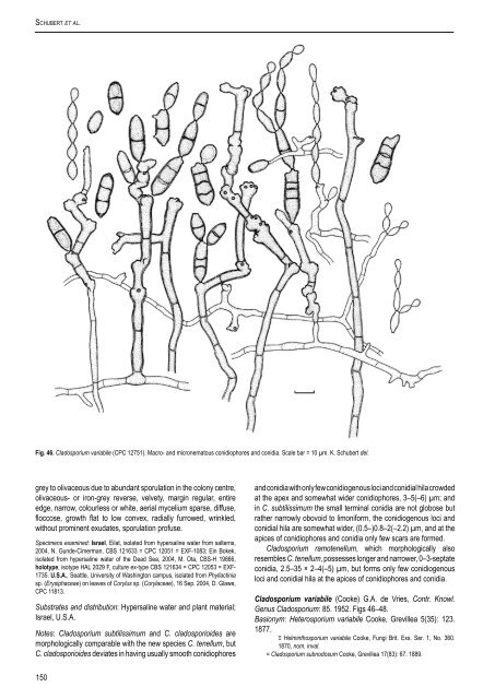
Schubert et al. Fig. 46. <strong>Cladosporium</strong> variabile (CPC 12751). Macro- <strong>and</strong> micronematous conidiophores <strong>and</strong> conidia. Scale bar = 10 µm. K. Schubert del. grey to olivaceous due to abundant sporulation in the colony centre, olivaceous- or iron-grey reverse, velvety, margin regular, entire edge, narrow, colourless or white, aerial mycelium sparse, diffuse, floccose, growth flat to low convex, radially furrowed, wrinkled, without prominent exudates, sporulation profuse. Specimens examined: Israel, Eilat, isolated from hypersaline water from salterns, 2004, N. Gunde-Cimerman, <strong>CBS</strong> 121633 = CPC 12051 = EXF-1083; Ein Bokek, isolated from hypersaline water of the Dead Sea, 2004, M. Ota, <strong>CBS</strong>-H 19866, holotype, isotype HAL 2029 F, culture ex-type <strong>CBS</strong> 121634 = CPC 12053 = EXF- 1735. U.S.A., Seattle, University of Washington campus, isolated from Phyllactinia sp. (Erysiphaceae) on leaves of Corylus sp. (Corylaceae), 16 Sep. 2004, D. Glawe, CPC 11813. Substrates <strong>and</strong> distribution: Hypersaline water <strong>and</strong> plant material; Israel, U.S.A. Notes: <strong>Cladosporium</strong> subtilissimum <strong>and</strong> C. cladosporioides are morphologically comparable with the new species C. tenellum, but C. cladosporioides deviates in having usually smooth conidiophores <strong>and</strong> conidia with only few conidiogenous loci <strong>and</strong> conidial hila crowded at the apex <strong>and</strong> somewhat wider conidiophores, 3–5(–6) µm; <strong>and</strong> in C. subtilissimum the small terminal conidia are not globose but rather narrowly obovoid to limoniform, the conidiogenous loci <strong>and</strong> conidial hila are somewhat wider, (0.5–)0.8–2(–2.2) µm, <strong>and</strong> at the apices of conidiophores <strong>and</strong> conidia only few scars are formed. <strong>Cladosporium</strong> ramotenellum, which morphologically also resembles C. tenellum, possesses longer <strong>and</strong> narrower, 0–3-septate conidia, 2.5–35 × 2–4(–5) µm, but forms only few conidiogenous loci <strong>and</strong> conidial hila at the apices of conidiophores <strong>and</strong> conidia. <strong>Cladosporium</strong> variabile (Cooke) G.A. de Vries, Contr. Knowl. Genus <strong>Cladosporium</strong>: 85. 1952. Figs 46–48. Basionym: Heterosporium variabile Cooke, Grevillea 5(35): 123. 1877. ≡ Helminthosporium variabile Cooke, Fungi Brit. Exs. Ser. 1, No. 360. 1870, nom. inval. = <strong>Cladosporium</strong> subnodosum Cooke, Grevillea 17(83): 67. 1889. 150
- Page 1:
Studies in Mycology 58 (2007) The g
- Page 4 and 5:
Studies in Mycology The Studies in
- Page 7 and 8:
CONTENTS P.W. Crous, U. Braun and J
- Page 9 and 10:
lectotype for the genus by Clements
- Page 11:
Schubert K (2005a). Morphotaxonomic
- Page 14 and 15:
Crous et al. Table 1. Isolates for
- Page 16 and 17:
Crous et al. Table 1. (Continued).
- Page 18 and 19:
Crous et al. 100 10 changes 65 100
- Page 20 and 21:
Crous et al. Treatment of phylogene
- Page 22 and 23:
Crous et al. Teratosphaeria bellula
- Page 24 and 25:
Crous et al. 6. Conidiophores short
- Page 26 and 27:
Crous et al. Habit plant pathogenic
- Page 28 and 29:
Crous et al. Fig. 7. Catenulostroma
- Page 30 and 31:
Crous et al. system, consisting of
- Page 32 and 33:
Crous et al. Fig. 9. Penidiella col
- Page 34 and 35:
Crous et al. Fig. 12. Penidiella ri
- Page 36 and 37:
Crous et al. conidiophores, about 1
- Page 38 and 39:
Crous et al. have conidiomata rangi
- Page 40 and 41:
Crous et al. Schizothyriaceae clade
- Page 42 and 43:
Crous et al. Fig. 21. Stigmidium sc
- Page 44 and 45:
Crous et al. Gams W, Verkley GJM, C
- Page 46 and 47:
Crous et al. Table 1. Isolates for
- Page 48 and 49:
Crous et al. 100 61 100 52 100 10 c
- Page 50 and 51:
Crous et al. Fig. 3. Rachicladospor
- Page 52 and 53:
Crous et al. Fig. 4. Toxicocladospo
- Page 54 and 55:
Crous et al. Fig. 5. Verrucocladosp
- Page 56 and 57:
Crous et al. Fig. 7. Stenella aragu
- Page 58 and 59:
Crous et al. Helotiales, incertae s
- Page 60 and 61:
Crous et al. Fig. 10. Ochrocladospo
- Page 62 and 63:
Crous et al. Fig. 12. Rhizocladospo
- Page 64 and 65:
Crous et al. 7. Conidiophores unbra
- Page 66 and 67:
Crous et al. 33. Terminal conidioge
- Page 68 and 69:
Crous et al. Crous PW, Kang JC, Bra
- Page 70 and 71:
Arzanlou et al. To date 26 species
- Page 72 and 73:
Arzanlou et al. Table 1. (Continued
- Page 74 and 75:
Arzanlou et al. 10 changes Athelia
- Page 76 and 77:
Arzanlou et al. Athelia epiphylla A
- Page 78 and 79:
Arzanlou et al. Fig. 3. Periconiell
- Page 80 and 81:
Arzanlou et al. Cultural characteri
- Page 82 and 83:
Arzanlou et al. Fig. 10. A. Ramichl
- Page 84 and 85:
Arzanlou et al. Ramichloridium musa
- Page 86 and 87:
Arzanlou et al. Cultural characteri
- Page 88 and 89:
Arzanlou et al. Fig. 20. Rhinocladi
- Page 90 and 91:
Arzanlou et al. Fig. 22. Rhinocladi
- Page 92 and 93:
Arzanlou et al. Rhinocladiella mack
- Page 94 and 95:
Arzanlou et al. Fig. 26. Veronaea c
- Page 96 and 97:
Arzanlou et al. Cultural characteri
- Page 98 and 99:
Arzanlou et al. Fig. 30. Myrmecridi
- Page 100 and 101:
Arzanlou et al. Fig. 32. Radulidium
- Page 102 and 103:
Arzanlou et al. Fig. 34. Rhodoveron
- Page 104 and 105:
Arzanlou et al. even further, thoug
- Page 106 and 107:
available online at www.studiesinmy
- Page 108 and 109:
Dichocladosporium gen. nov. 10 chan
- Page 110 and 111: Dichocladosporium gen. nov. Fig. 3.
- Page 112 and 113: Dichocladosporium gen. nov. to intr
- Page 114 and 115: Dichocladosporium gen. nov. Czechos
- Page 116 and 117: available online at www.studiesinmy
- Page 118 and 119: Cladosporium herbarum species compl
- Page 120 and 121: Cladosporium herbarum species compl
- Page 122 and 123: Cladosporium herbarum species compl
- Page 124 and 125: Cladosporium herbarum species compl
- Page 126 and 127: Cladosporium herbarum species compl
- Page 128 and 129: Cladosporium herbarum species compl
- Page 130 and 131: Cladosporium herbarum species compl
- Page 132 and 133: Cladosporium herbarum species compl
- Page 134 and 135: Cladosporium herbarum species compl
- Page 136 and 137: Cladosporium herbarum species compl
- Page 138 and 139: Cladosporium herbarum species compl
- Page 140 and 141: Cladosporium herbarum species compl
- Page 142 and 143: Cladosporium herbarum species compl
- Page 144 and 145: Cladosporium herbarum species compl
- Page 146 and 147: Cladosporium herbarum species compl
- Page 148 and 149: Cladosporium herbarum species compl
- Page 150 and 151: Cladosporium herbarum species compl
- Page 152 and 153: Cladosporium herbarum species compl
- Page 154 and 155: Cladosporium herbarum species compl
- Page 156 and 157: Cladosporium herbarum species compl
- Page 158 and 159: Cladosporium herbarum species compl
- Page 162 and 163: Cladosporium herbarum species compl
- Page 164 and 165: Cladosporium herbarum species compl
- Page 166 and 167: Cladosporium herbarum species compl
- Page 168 and 169: available online at www.studiesinmy
- Page 170 and 171: Cladosporium sphaerospermum species
- Page 172 and 173: Cladosporium sphaerospermum species
- Page 174 and 175: Cladosporium sphaerospermum species
- Page 176 and 177: Cladosporium sphaerospermum species
- Page 178 and 179: Cladosporium sphaerospermum species
- Page 180 and 181: Cladosporium sphaerospermum species
- Page 182 and 183: Cladosporium sphaerospermum species
- Page 184 and 185: Cladosporium sphaerospermum species
- Page 186 and 187: Cladosporium sphaerospermum species
- Page 188 and 189: Cladosporium sphaerospermum species
- Page 190 and 191: Cladosporium sphaerospermum species
- Page 192 and 193: Cladosporium sphaerospermum species
- Page 194 and 195: Cladosporium sphaerospermum species
- Page 196 and 197: Crous et al. The aim of the present
- Page 198 and 199: Crous et al. 10 changes Athelia epi
- Page 200 and 201: Crous et al. Table 1. Isolates used
- Page 202 and 203: Crous et al. Table 1. (Continued).
- Page 204 and 205: Crous et al. 0.1 expected changes p
- Page 206 and 207: Crous et al. Fig. 6. Cladophialopho
- Page 208 and 209: Crous et al. Fig. 11. Cladophialoph
- Page 210 and 211:
Crous et al. Fig. 13. Cladophialoph
- Page 212 and 213:
Crous et al. Fig. 15. Exophiala sp.
- Page 214 and 215:
Crous et al. Fig. 18. Cylindrosympo
- Page 216 and 217:
Crous et al. Fig. 19. Fusicladium a
- Page 218 and 219:
Crous et al. Fig. 22. Fusicladium f
- Page 220 and 221:
Crous et al. Fig. 24. Fusicladium p
- Page 222 and 223:
Crous et al. Fusicladium rhodense C
- Page 224 and 225:
Crous et al. Excluded taxa Polyscyt
- Page 226 and 227:
Crous et al. the conidial tips are
- Page 228 and 229:
available online at www.studiesinmy
- Page 230 and 231:
Cladophialophora carrionii complex
- Page 232 and 233:
Cladophialophora carrionii complex
- Page 234 and 235:
Cladophialophora carrionii complex
- Page 236 and 237:
Cladophialophora carrionii complex
- Page 238 and 239:
Cladophialophora carrionii complex
- Page 240 and 241:
Cladophialophora carrionii complex
- Page 242 and 243:
Cladophialophora carrionii complex
- Page 244 and 245:
available online at www.studiesinmy
- Page 246 and 247:
Hormoconis resinae and morphologica
- Page 248 and 249:
Hormoconis resinae and morphologica
- Page 250 and 251:
Hormoconis resinae and morphologica
- Page 252 and 253:
Fig. 6 Hormoconis resinae and morph
- Page 254 and 255:
Hormoconis resinae and morphologica
- Page 256 and 257:
Cladophialophora, 52 k , 54 k -55,
- Page 258 and 259:
Fusicladium phillyreae, 189 c , 191
- Page 260 and 261:
Ramichloridium epichloës, 60, 89 P
- Page 262:
Trimmatostroma salicis, 3 t , 5, 6