

The genus Cladosporium and similar dematiaceous ... - CBS - KNAW
The genus Cladosporium and similar dematiaceous ... - CBS - KNAW
The genus Cladosporium and similar dematiaceous ... - CBS - KNAW

You also want an ePaper? Increase the reach of your titles
YUMPU automatically turns print PDFs into web optimized ePapers that Google loves.
Schubert et al.<br />
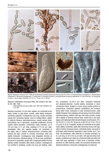
Fig. 18. <strong>Cladosporium</strong> herbarum (CPC 11600) <strong>and</strong> its teleomorph Davidiella tassiana (from the host <strong>and</strong> CPC 12181). A–B. Macronematous conidiophores. C. Micronematous<br />
conidiophore. D. Microcyclic conidiogenesis. E. Conidial chain. F. Ascomata on the leaf. G. Ascomata formed in culture on nettle stems. H–I. Asci on the host. J–K. Ascospores<br />
in culture. L. Asci in culture. Scale bars: A, E, H, J–L = 10 µm, F–G, I = 200 µm.<br />
Basionym: Didymellina macrospora Kleb., Ber. Deutsch. Bot. Ges.<br />
42: 60, 1924. 1925.<br />
≡ Mycosphaerella macrospora (Kleb.) Jørst., Meld. Stat. Plantepatol. Inst.<br />
1: 20. 1945.<br />
Mycelium branched, 2–8 µm wide, septate, not constricted at the<br />
septa, hyaline to pale brown, smooth, walls slightly thickened,<br />
sometimes guttulate. Conidiophores very long, usually terminally<br />
arising from ascending hyphae, erect to subdecumbent, slightly<br />
to distinctly flexuous, geniculate-sinuous, usually several times,<br />
subnodulose due to geniculate, sympodial proliferation forming<br />
swollen lateral shoulders, unbranched, rarely branched, up to<br />
720 µm long, 6–11 µm wide, swellings 8–11(–14) µm wide,<br />
pluriseptate, often very regularly septate, not constricted at<br />
the septa, pale to medium olivaceous-brown, somewhat paler<br />
towards the apex, smooth to minutely verruculose, walls only<br />
slightly thickened. Conidiogenous cells integrated, terminal as<br />
well as intercalary, cylindrical-oblong, 15–55 µm long, proliferation<br />
percurrent to sympodial, usually with a single geniculation forming<br />
laterally swollen shoulders often below a septum, conidiogenous<br />
loci confined to swellings, usually one locus per swelling, rarely<br />
two, protuberant, (2–)2.5–4 µm diam, somewhat thickened<br />
<strong>and</strong> darkened-refractive. Conidia solitary, sometimes in short,<br />
unbranched chains, straight to curved, young conidia pyriform to<br />
subcylindrical, connection between conidiophore <strong>and</strong> conidium<br />
being rather broad, subhyaline to pale olivaceous-brown, walls<br />
slightly thickened, then enlarging <strong>and</strong> becoming more thick-walled,<br />
cylindrical-oblong, soleiform with age, both ends rounded, usually<br />
with a slightly to distinctly bulbous base, visible from a very early<br />
stage, but broadest part often towards the apex not at the base, (18–)<br />
30–75(–87) × (7–)10–16(–18) µm [av. ± SD, 53.3 (± 17.8) × 12.6 (±<br />
2.2) µm], (0–)2–6(–7)-septate, usually not constricted at the septa,<br />
rarely slightly constricted, septa often becoming sinuous with age,<br />
pale to medium olivaceous-brown, sometimes darker, verrucose to<br />
echinulate, walls thickened, especially in older conidia, up to 1 µm<br />
thick, hila protuberant, often stalk-like or conically prolonged, up<br />
to 2 µm long, (2–)2.5–3.5(–4) µm diam, with age becoming more<br />
sessile, sometimes just visible as a thickened plate just below the<br />
outer wall layer, especially in distal scars of branched conidia,<br />
periclinal rim often distinctly visible, hila somewhat thickened <strong>and</strong><br />
darkened-refractive; microcyclic conidiogenesis not observed.<br />
126


