

The genus Cladosporium and similar dematiaceous ... - CBS - KNAW
The genus Cladosporium and similar dematiaceous ... - CBS - KNAW
The genus Cladosporium and similar dematiaceous ... - CBS - KNAW

Create successful ePaper yourself
Turn your PDF publications into a flip-book with our unique Google optimized e-Paper software.
Crous et al.<br />
conidiophores, about 10–15 × 2–3 µm. Conidiogenous cells<br />
terminal <strong>and</strong> intercalary, unbranched, subcylindrical, 5–12 × 3–4<br />
µm, medium brown, smooth or almost so to finely verruculose,<br />
apex of conidiogenous cells frequently swollen, up to 6 µm diam,<br />
with 1–3(–4) flat-tipped, non to slightly thickened, non to slightly<br />
darkened-refractive loci, 1–1.5 µm wide, frequently appearing<br />
subdenticulate, up to 1.5 µm long, intercalary conidiogenous cells<br />
also slightly swollen at the conidiogenous portion just below the<br />
upper septum, which render the conidiophores subnodulose to<br />
nodulose, swellings round about the conidiophore axis or unilateral.<br />
Conidia ellipsoid-ovoid, subcylindrical, pale to medium olivaceousbrown<br />
or brown, finely verruculose, wall ≤ 0.5 µm wide, guttulate or<br />
not, occurring in branched chains. Ramoconidia 0–1(–3)-septate,<br />
5–15(–22) × 3–4(–5) µm, with 1–3 subdenticulate apical hila;<br />
secondary conidia 0(–1)-septate, ellipsoid, obovoid to irregular,<br />
(4–)5–7(–8) × (2–)2.5–3(–4) µm; hila non to slightly thickened, non<br />
to slightly darkened-refractive, (0.5–)1(–1.5) µm wide.<br />
Cultural characteristics: Colonies on OA erumpent, spreading,<br />
with dense, compact aerial mycelium, <strong>and</strong> even, smooth margins;<br />
olivaceous-grey (surface), margins iron-grey. Colonies reaching 22<br />
mm diam after 1 mo at 25 °C in the dark.<br />
Specimen examined: Venezuela, isolated from man with tinea nigra, Jan. 1975, D.<br />
Borelli, holotype <strong>CBS</strong> H-19934, culture ex-type <strong>CBS</strong> 106.75.<br />
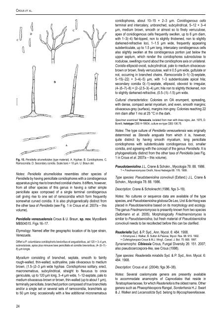
Fig. 15. Penidiella strumelloidea (type material). A. Hyphae. B. Conidiophores. C.<br />
Ramoconidia. D. Secondary conidia. Scale bars = 10 µm. U. Braun del.<br />
Notes: Penidiella strumelloidea resembles other species of<br />
Penidiella by having penicillate conidiophores with a conidiogenous<br />
apparatus giving rise to branched conidial chains. It differs, however,<br />
from all other species of this <strong>genus</strong> in having a rather simple<br />
penicillate apex composed of a single terminal conidiogenous<br />
cell giving rise to one set of ramoconidia which form frequently<br />
somewhat curved conidia. It is also phylogenetically distinct from<br />
the other taxa of Penidiella (see Fig. 1 in Crous et al. 2007a – this<br />
volume).<br />
Penidiella venezuelensis Crous & U. Braun, sp. nov. MycoBank<br />
MB504515. Figs 16–17.<br />
Etymology: Named after the geographic location of its type strain,<br />
Venezuela.<br />
Differt a P. columbiana conidiophoris bevioribus et angustioribus, ad 120 × 3–4 µm,<br />
subnodulosis, apice plus minusve laxe penicillatis et conidiis brevioribus, (4–)5–7(–<br />
8) µm longis.<br />
Mycelium consisting of branched, septate, smooth to faintly<br />
rough-walled, thin-walled, subhyaline, pale olivaceous to medium<br />
brown, (1.5–)2–3 µm wide hyphae. Conidiophores solitary, erect,<br />
macronematous, subcylindrical, straight to flexuous to once<br />
geniculate, up to 120 µm long, 3–4 µm wide, 1–12-septate, pale to<br />
medium olivaceous-brown or brown, thin-walled (up to about 1 µm),<br />
terminally penicillate, branched portion composed of true branchlets<br />
<strong>and</strong>/or a single set or several sets of ramoconidia, branchlets up<br />
to 50 µm long; occasionally with a few additional micronematous<br />
Notes: <strong>The</strong> type culture of Penidiella venezuelensis was originally<br />
determined as Stenella araguata from which it is, however,<br />
quite distinct by having smooth mycelium, long penicillate<br />
conidiophores with subdenticulate conidiogenous loci, smaller<br />
conidia, <strong>and</strong> agreeing with the concept of the <strong>genus</strong> Penidiella. It is<br />
phylogenetically distinct from the other taxa of Penidiella (see Fig.<br />
1 in Crous et al. 2007a – this volume).<br />
Pseudotaeniolina J.L. Crane & Schokn., Mycologia 78: 88. 1986.<br />
? = Friedmanniomyces Onofri, Nova Hedwigia 68: 176. 1999.<br />
Type species: Pseudotaeniolina convolvuli (Esf<strong>and</strong>.) J.L. Crane &<br />
Schokn., Mycologia 78: 88. 1986.<br />
Description: Crane & Schoknecht (1986, figs 3–19).<br />
Notes: No cultures or sequence data are available of the type<br />
species, <strong>and</strong> Pseudotaeniolina globosa De Leo, Urzì & de Hoog was<br />
placed in Pseudotaeniolina based on its morphology <strong>and</strong> ecology.<br />
<strong>The</strong> <strong>genus</strong> Friedmanniomyces is presently known from two species<br />
(Selbmann et al. 2005). Morphologically Friedmanniomyces is<br />
<strong>similar</strong> to Pseudotaeniolina, but fresh material of Pseudotaeniolina<br />
convolvuli needs to be recollected before this can be clarified.<br />
Readeriella Syd. & P. Syd., Ann. Mycol. 6: 484. 1908.<br />
= Kirramyces J. Walker, B. Sutton & Pascoe, Mycol. Res. 96: 919. 1992.<br />
= Colletogloeopsis Crous & M.J. Wingf., Canad. J. Bot. 75: 668. 1997.<br />
Synanamorphs: Cibiessia Crous, Fungal Diversity 26: 151. 2007;<br />
also pseudocercospora-like, see Crous (1998).<br />
Type species: Readeriella mirabilis Syd. & P. Syd., Ann. Mycol. 6:<br />
484. 1908.<br />
Description: Crous et al. (2004b; figs 36–38).<br />
Notes: Several coelomycete genera are presently available<br />
to accommodate anamorphs of Capnodiales that reside in<br />
Teratosphaeriaceae, for which Readeriella is the oldest name. Other<br />
genera such as Phaeophleospora Rangel, Sonderhenia H.J. Swart<br />
& J. Walker <strong>and</strong> Lecanosticta Syd. belong to Mycosphaerellaceae.<br />
24


