Arzanlou et al. Cultural characteristics: Colonies on MEA reaching 12 mm diam after 14 d at 24 °C, with entire, smooth, sharp margin; mycelium compacted, becoming hairy, colonies up to 1 mm high; surface olivaceous to olivaceous-grey, reverse dark grey-olivaceous to olivaceous-black. Specimen examined: South Africa, Western Cape Province, Kogelberg, on dead culms of Ischyrolepis subverticillata, May 2001, S. Lee, holotype <strong>CBS</strong> H-19927, culture ex-type <strong>CBS</strong> 113477. Periconiella levispora Arzanlou, W. Gams & Crous, sp. nov. MycoBank MB504546. Figs 5–6B. Etymology: (Latin) levis = smooth. A simili Periconiella velutina conidiis levibus et maioribus, ad 23 μm longis distinguenda. In vitro: Submerged hyphae smooth, hyaline, thin-walled, 2–2.5 µm wide; aerial hyphae subhyaline, later becoming dark brown, thickwalled, smooth. Conidiophores arising vertically from creeping aerial hyphae, dark brown at the base, paler towards the apex, thick-walled; in the upper part bearing several short branches, up to 120 µm long. Conidiogenous cells integrated, occasionally discrete, cylindrical, variable in length, 10–20 µm long, subhyaline, later becoming pale brown, fertile part as wide as the basal part, proliferating sympodially, forming a short rachis with pigmented <strong>and</strong> slightly thickened, somewhat protruding scars, less than 1 µm diam. Conidia solitary, 0(–2)-septate, smooth, pale olivaceous, cylindrical, ellipsoidal, pyriform to clavate, (7–)11–14(–23) × (3–)4– 5(–6) µm, with a truncate base <strong>and</strong> a darkened, slightly thickened hilum, 2 µm diam. Cultural characteristics: Colonies on MEA slow-growing, reaching 5 mm diam after 14 d at 24 °C, with entire margin; aerial mycelium compact, raised, velvety, olivaceous-grey; reverse olivaceousblack. Basionym: Chloridium apiculatum J.H. Mill., Giddens & A.A. Foster, Mycologia 49: 789. 1957. ≡ Veronaea apiculata (J.H. Mill., Giddens & A.A. Foster) M.B. Ellis, in Ellis, More Dematiaceous Hyphomycetes: 209. 1976. [non Rhinocladiella apiculata Matsush., in Matsushima, Icon. Microfung. Mats. lect.: 122. 1975]. = Rhinocladiella indica Agarwal, Lloydia 32: 388. 1969. [non Chloridium indicum Subram., Proc. Indian Acad. Sci., Sect. B, 42: 286. 1955]. In vitro: Submerged hyphae hyaline to subhyaline, thin-walled, 1–2.5 µm wide; aerial hyphae slightly darker, smooth-walled. Conidiophores generally arising at right angles from creeping aerial hyphae, straight, unbranched, thick-walled, dark brown, continuous or with 1–2(–3) additional thin septa, up to 100 µm long; intercalary cells 10–28 µm long. Conidiogenous cells integrated, terminal, smooth, thick-walled, golden-brown, straight, cylindrical, 25–37(– 47) × 2–3.5 µm; proliferating sympodially, resulting in a straight rachis with conspicuous conidiogenous loci; scars prominent, crowded, slightly pigmented, less than 1 µm diam. Conidia solitary, obovate to obconical, pale brown, finely verrucose, (3–)5–5.5(–7.5) × (2–)2.5–3(–4) µm, hilum conspicuous, slightly pigmented, about 1 µm diam. Cultural characteristics: Colonies on MEA reaching 35 mm diam after 14 d at 24 °C; minimum temperature for growth above 6 °C, optimum 24 °C, maximum 30 °C. Colonies raised, velvety, dense, with entire margin; surface olivaceous-green, reverse olivaceousblack, often with a diffusing citron-yellow pigment. Specimens examined: Pakistan, Lahore, from soil, A. Kamal, <strong>CBS</strong> 400.76 = IMI 088021. South Africa, from preserved Cucumis sativus in 8-oxyquinoline sulphate, M.C. Papendorf, <strong>CBS</strong> 390.67; Potchefstroom, from Aloe sp., M.C. Papendorf, <strong>CBS</strong> 391.67. U.S.A., Georgia, isolated from forest soil, <strong>CBS</strong> 156.59 = ATCC 13211 = IMI 100716 = QM 7716, ex-type culture. Specimen examined: Sri Lanka, Hakgala Botanic Gardens, on dead leaves of Turpinia pomifera, Jan. 1973, W. Gams, holotype <strong>CBS</strong> H-15611, culture ex-type <strong>CBS</strong> 873.73. Ramichloridium Stahel ex de Hoog, Stud. Mycol. 15: 59. 1977. In vitro: Colonies flat to raised, with entire margin; surface olivaceous-green to olivaceous-black. Mycelium consisting of submerged <strong>and</strong> aerial hyphae; submerged hyphae hyaline to subhyaline, thin-walled, aerial hyphae smooth or verrucose. Conidiophores straight, unbranched, rarely branched, thick-walled, dark brown (darker than the subtending hyphae), continuous or with several additional thin septa. Conidiogenous cells integrated, terminal, polyblastic, smooth, thick-walled, golden-brown, apical part subhyaline, with sympodial proliferation, straight or flexuose, geniculate or nodose, with conspicuous conidiogenous loci; scars crowded or scattered, unthickened, unpigmented to faintly pigmented, or slightly prominent denticles. Conidia solitary, 0–1- septate, subhyaline to pale brown, smooth to coarsely verrucose, rather thin-walled, obovate, obconical or globose to ellipsoidal, fusiform, with a somewhat prominent, slightly pigmented hilum; conidial secession schizolytic. Type species: R. apiculatum (J.H. Mill., Giddens & A.A. Foster) de Hoog, Stud. Mycol. 15: 69. 1977. Ramichloridium apiculatum (J.H. Mill., Giddens & A.A. Foster) de Hoog, Stud. Mycol. 15: 69. 1977. Fig. 8. Fig. 7. A. Periconiella arcuata (<strong>CBS</strong> 113477). B. Myrmecridium schulzeri (<strong>CBS</strong> 325.74). C. Thysanorea papuana (<strong>CBS</strong> 212.96). Scale bars = 10 µm. 68
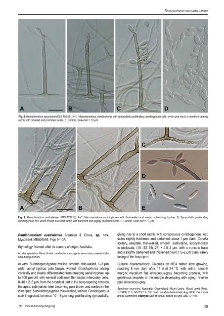
Ramichloridium <strong>and</strong> allied genera Fig. 8. Ramichloridium apiculatum (<strong>CBS</strong> 156.59). A–C. Macronematous conidiophores with sympodially proliferating conidiogenous cells, which give rise to a conidium-bearing rachis with crowded <strong>and</strong> prominent scars. D. Conidia. Scale bar = 10 µm. Fig. 9. Ramichloridium australiense (<strong>CBS</strong> 121710). A–C. Macronematous conidiophores with thick-walled <strong>and</strong> warted subtending hyphae. D. Sympodially proliferating conidiogenous cell, which results in a short rachis with darkened <strong>and</strong> slightly thickened scars. E. Conidia. Scale bar = 10 µm. Ramichloridium australiense Arzanlou & Crous, sp. nov. MycoBank MB504548. Figs 9–10A. Etymology: Named after its country of origin, Australia. Ab aliis speciebus Ramichloridii conidiophoris ex hyphis verrucosis, crassitunicatis ortis distinguendum. In vitro: Submerged hyphae hyaline, smooth, thin-walled, 1–2 µm wide; aerial hyphae pale brown, warted. Conidiophores arising vertically <strong>and</strong> clearly differentiated from creeping aerial hyphae, up to 400 µm tall, with several additional thin septa; intercalary cells, 8–40 × 2–5 µm, from the broadest part at the base tapering towards the apex, subhyaline, later becoming pale brown <strong>and</strong> warted in the lower part. Subtending hyphae thick-walled, warted. Conidiogenous cells integrated, terminal, 10–18 µm long, proliferating sympodially, www.studiesinmycology.org giving rise to a short rachis with conspicuous conidiogenous loci; scars slightly thickened <strong>and</strong> darkened, about 1 µm diam. Conidia solitary, aseptate, thin-walled, smooth, subhyaline, subcylindrical to obclavate, (10–)12–15(–23) × 2.5–3 µm, with a truncate base <strong>and</strong> a slightly darkened <strong>and</strong> thickened hilum,1.5–2 µm diam, rarely fusing at the basal part. Cultural characteristics: Colonies on MEA rather slow growing, reaching 8 mm diam after 14 d at 24 °C, with entire, smooth margin; mycelium flat, olivaceous-grey, becoming granular, with gelatinous droplets at the margin developing with aging; reverse pale olivaceous-grey. Specimen examined: Australia, Queensl<strong>and</strong>, Mount Lewis, Mount Lewis Road, 16°34’47.2” S, 145°19’7” E, 538 m alt., on Musa banksii leaf, Aug. 2006, P.W. Crous <strong>and</strong> B. Summerell, holotype <strong>CBS</strong> H-19928, culture ex-type <strong>CBS</strong> 121710. 69
- Page 1:
Studies in Mycology 58 (2007) The g
- Page 4 and 5:
Studies in Mycology The Studies in
- Page 7 and 8:
CONTENTS P.W. Crous, U. Braun and J
- Page 9 and 10:
lectotype for the genus by Clements
- Page 11:
Schubert K (2005a). Morphotaxonomic
- Page 14 and 15:
Crous et al. Table 1. Isolates for
- Page 16 and 17:
Crous et al. Table 1. (Continued).
- Page 18 and 19:
Crous et al. 100 10 changes 65 100
- Page 20 and 21:
Crous et al. Treatment of phylogene
- Page 22 and 23:
Crous et al. Teratosphaeria bellula
- Page 24 and 25:
Crous et al. 6. Conidiophores short
- Page 26 and 27:
Crous et al. Habit plant pathogenic
- Page 28 and 29:
Crous et al. Fig. 7. Catenulostroma
- Page 30 and 31: Crous et al. system, consisting of
- Page 32 and 33: Crous et al. Fig. 9. Penidiella col
- Page 34 and 35: Crous et al. Fig. 12. Penidiella ri
- Page 36 and 37: Crous et al. conidiophores, about 1
- Page 38 and 39: Crous et al. have conidiomata rangi
- Page 40 and 41: Crous et al. Schizothyriaceae clade
- Page 42 and 43: Crous et al. Fig. 21. Stigmidium sc
- Page 44 and 45: Crous et al. Gams W, Verkley GJM, C
- Page 46 and 47: Crous et al. Table 1. Isolates for
- Page 48 and 49: Crous et al. 100 61 100 52 100 10 c
- Page 50 and 51: Crous et al. Fig. 3. Rachicladospor
- Page 52 and 53: Crous et al. Fig. 4. Toxicocladospo
- Page 54 and 55: Crous et al. Fig. 5. Verrucocladosp
- Page 56 and 57: Crous et al. Fig. 7. Stenella aragu
- Page 58 and 59: Crous et al. Helotiales, incertae s
- Page 60 and 61: Crous et al. Fig. 10. Ochrocladospo
- Page 62 and 63: Crous et al. Fig. 12. Rhizocladospo
- Page 64 and 65: Crous et al. 7. Conidiophores unbra
- Page 66 and 67: Crous et al. 33. Terminal conidioge
- Page 68 and 69: Crous et al. Crous PW, Kang JC, Bra
- Page 70 and 71: Arzanlou et al. To date 26 species
- Page 72 and 73: Arzanlou et al. Table 1. (Continued
- Page 74 and 75: Arzanlou et al. 10 changes Athelia
- Page 76 and 77: Arzanlou et al. Athelia epiphylla A
- Page 78 and 79: Arzanlou et al. Fig. 3. Periconiell
- Page 82 and 83: Arzanlou et al. Fig. 10. A. Ramichl
- Page 84 and 85: Arzanlou et al. Ramichloridium musa
- Page 86 and 87: Arzanlou et al. Cultural characteri
- Page 88 and 89: Arzanlou et al. Fig. 20. Rhinocladi
- Page 90 and 91: Arzanlou et al. Fig. 22. Rhinocladi
- Page 92 and 93: Arzanlou et al. Rhinocladiella mack
- Page 94 and 95: Arzanlou et al. Fig. 26. Veronaea c
- Page 96 and 97: Arzanlou et al. Cultural characteri
- Page 98 and 99: Arzanlou et al. Fig. 30. Myrmecridi
- Page 100 and 101: Arzanlou et al. Fig. 32. Radulidium
- Page 102 and 103: Arzanlou et al. Fig. 34. Rhodoveron
- Page 104 and 105: Arzanlou et al. even further, thoug
- Page 106 and 107: available online at www.studiesinmy
- Page 108 and 109: Dichocladosporium gen. nov. 10 chan
- Page 110 and 111: Dichocladosporium gen. nov. Fig. 3.
- Page 112 and 113: Dichocladosporium gen. nov. to intr
- Page 114 and 115: Dichocladosporium gen. nov. Czechos
- Page 116 and 117: available online at www.studiesinmy
- Page 118 and 119: Cladosporium herbarum species compl
- Page 120 and 121: Cladosporium herbarum species compl
- Page 122 and 123: Cladosporium herbarum species compl
- Page 124 and 125: Cladosporium herbarum species compl
- Page 126 and 127: Cladosporium herbarum species compl
- Page 128 and 129: Cladosporium herbarum species compl
- Page 130 and 131:
Cladosporium herbarum species compl
- Page 132 and 133:
Cladosporium herbarum species compl
- Page 134 and 135:
Cladosporium herbarum species compl
- Page 136 and 137:
Cladosporium herbarum species compl
- Page 138 and 139:
Cladosporium herbarum species compl
- Page 140 and 141:
Cladosporium herbarum species compl
- Page 142 and 143:
Cladosporium herbarum species compl
- Page 144 and 145:
Cladosporium herbarum species compl
- Page 146 and 147:
Cladosporium herbarum species compl
- Page 148 and 149:
Cladosporium herbarum species compl
- Page 150 and 151:
Cladosporium herbarum species compl
- Page 152 and 153:
Cladosporium herbarum species compl
- Page 154 and 155:
Cladosporium herbarum species compl
- Page 156 and 157:
Cladosporium herbarum species compl
- Page 158 and 159:
Cladosporium herbarum species compl
- Page 160 and 161:
Cladosporium herbarum species compl
- Page 162 and 163:
Cladosporium herbarum species compl
- Page 164 and 165:
Cladosporium herbarum species compl
- Page 166 and 167:
Cladosporium herbarum species compl
- Page 168 and 169:
available online at www.studiesinmy
- Page 170 and 171:
Cladosporium sphaerospermum species
- Page 172 and 173:
Cladosporium sphaerospermum species
- Page 174 and 175:
Cladosporium sphaerospermum species
- Page 176 and 177:
Cladosporium sphaerospermum species
- Page 178 and 179:
Cladosporium sphaerospermum species
- Page 180 and 181:
Cladosporium sphaerospermum species
- Page 182 and 183:
Cladosporium sphaerospermum species
- Page 184 and 185:
Cladosporium sphaerospermum species
- Page 186 and 187:
Cladosporium sphaerospermum species
- Page 188 and 189:
Cladosporium sphaerospermum species
- Page 190 and 191:
Cladosporium sphaerospermum species
- Page 192 and 193:
Cladosporium sphaerospermum species
- Page 194 and 195:
Cladosporium sphaerospermum species
- Page 196 and 197:
Crous et al. The aim of the present
- Page 198 and 199:
Crous et al. 10 changes Athelia epi
- Page 200 and 201:
Crous et al. Table 1. Isolates used
- Page 202 and 203:
Crous et al. Table 1. (Continued).
- Page 204 and 205:
Crous et al. 0.1 expected changes p
- Page 206 and 207:
Crous et al. Fig. 6. Cladophialopho
- Page 208 and 209:
Crous et al. Fig. 11. Cladophialoph
- Page 210 and 211:
Crous et al. Fig. 13. Cladophialoph
- Page 212 and 213:
Crous et al. Fig. 15. Exophiala sp.
- Page 214 and 215:
Crous et al. Fig. 18. Cylindrosympo
- Page 216 and 217:
Crous et al. Fig. 19. Fusicladium a
- Page 218 and 219:
Crous et al. Fig. 22. Fusicladium f
- Page 220 and 221:
Crous et al. Fig. 24. Fusicladium p
- Page 222 and 223:
Crous et al. Fusicladium rhodense C
- Page 224 and 225:
Crous et al. Excluded taxa Polyscyt
- Page 226 and 227:
Crous et al. the conidial tips are
- Page 228 and 229:
available online at www.studiesinmy
- Page 230 and 231:
Cladophialophora carrionii complex
- Page 232 and 233:
Cladophialophora carrionii complex
- Page 234 and 235:
Cladophialophora carrionii complex
- Page 236 and 237:
Cladophialophora carrionii complex
- Page 238 and 239:
Cladophialophora carrionii complex
- Page 240 and 241:
Cladophialophora carrionii complex
- Page 242 and 243:
Cladophialophora carrionii complex
- Page 244 and 245:
available online at www.studiesinmy
- Page 246 and 247:
Hormoconis resinae and morphologica
- Page 248 and 249:
Hormoconis resinae and morphologica
- Page 250 and 251:
Hormoconis resinae and morphologica
- Page 252 and 253:
Fig. 6 Hormoconis resinae and morph
- Page 254 and 255:
Hormoconis resinae and morphologica
- Page 256 and 257:
Cladophialophora, 52 k , 54 k -55,
- Page 258 and 259:
Fusicladium phillyreae, 189 c , 191
- Page 260 and 261:
Ramichloridium epichloës, 60, 89 P
- Page 262:
Trimmatostroma salicis, 3 t , 5, 6