Properties of hemp fibre polymer composites -An optimisation of ...
Properties of hemp fibre polymer composites -An optimisation of ...
Properties of hemp fibre polymer composites -An optimisation of ...

Create successful ePaper yourself
Turn your PDF publications into a flip-book with our unique Google optimized e-Paper software.
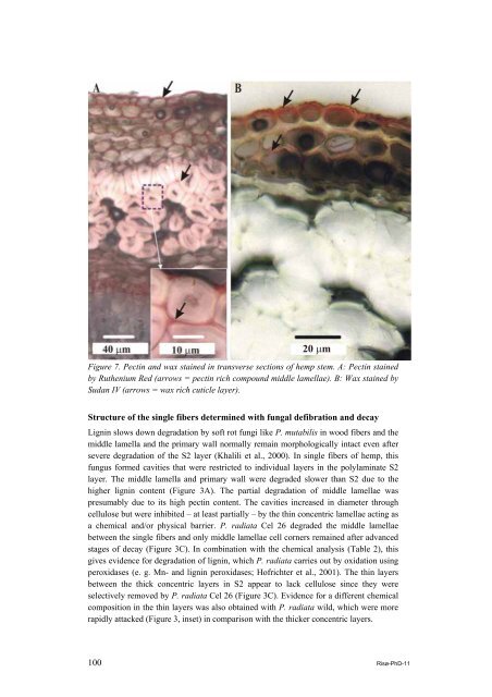
Figure 7. Pectin and wax stained in transverse sections <strong>of</strong> <strong>hemp</strong> stem. A: Pectin stained<br />
by Ruthenium Red (arrows = pectin rich compound middle lamellae). B: Wax stained by<br />
Sudan IV (arrows = wax rich cuticle layer).<br />
Structure <strong>of</strong> the single fibers determined with fungal defibration and decay<br />
Lignin slows down degradation by s<strong>of</strong>t rot fungi like P. mutabilis in wood fibers and the<br />
middle lamella and the primary wall normally remain morphologically intact even after<br />
severe degradation <strong>of</strong> the S2 layer (Khalili et al., 2000). In single fibers <strong>of</strong> <strong>hemp</strong>, this<br />
fungus formed cavities that were restricted to individual layers in the polylaminate S2<br />
layer. The middle lamella and primary wall were degraded slower than S2 due to the<br />
higher lignin content (Figure 3A). The partial degradation <strong>of</strong> middle lamellae was<br />
presumably due to its high pectin content. The cavities increased in diameter through<br />
cellulose but were inhibited – at least partially – by the thin concentric lamellae acting as<br />
a chemical and/or physical barrier. P. radiata Cel 26 degraded the middle lamellae<br />
between the single fibers and only middle lamellae cell corners remained after advanced<br />
stages <strong>of</strong> decay (Figure 3C). In combination with the chemical analysis (Table 2), this<br />
gives evidence for degradation <strong>of</strong> lignin, which P. radiata carries out by oxidation using<br />
peroxidases (e. g. Mn- and lignin peroxidases; H<strong>of</strong>richter et al., 2001). The thin layers<br />
between the thick concentric layers in S2 appear to lack cellulose since they were<br />
selectively removed by P. radiata Cel 26 (Figure 3C). Evidence for a different chemical<br />
composition in the thin layers was also obtained with P. radiata wild, which were more<br />
rapidly attacked (Figure 3, inset) in comparison with the thicker concentric layers.<br />
100 Risø-PhD-11