Properties of hemp fibre polymer composites -An optimisation of ...
Properties of hemp fibre polymer composites -An optimisation of ...
Properties of hemp fibre polymer composites -An optimisation of ...

You also want an ePaper? Increase the reach of your titles
YUMPU automatically turns print PDFs into web optimized ePapers that Google loves.
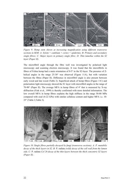
Figure 9. Hemp stem shown at increasing magnification using different transverse<br />
sections in SEM: A: Xylem + cambium + cortex + epidermis; B: Primary and secondary<br />
single <strong>fibre</strong>s; C: Major layers in primary single <strong>fibre</strong>; D: Thin lamellae within the S2<br />
layer (Paper II).<br />
The micr<strong>of</strong>ibril angle through the <strong>fibre</strong> wall was investigated by polarised light<br />
microscopy and scanning electron microscopy. It was found that the micr<strong>of</strong>ibrils in<br />
<strong>fibre</strong>s <strong>of</strong> Felina <strong>hemp</strong> had a main orientation <strong>of</strong> 0-5° in the S2-layer. The presence <strong>of</strong> Zhelical<br />
angles in the range 25-30° was observed (Figure 11A), but with variation<br />
between the <strong>fibre</strong>s (Paper II). Difference in micr<strong>of</strong>ibril angle is also present between<br />
early wood and late wood (Table 2). Superficial attack <strong>of</strong> <strong>hemp</strong> <strong>fibre</strong>s (Figure 11C) and<br />
polarization light microscopy showed the S1 layer with micr<strong>of</strong>ibril angles in the range <strong>of</strong><br />
70-90° (Paper II). The average MFA in <strong>hemp</strong> <strong>fibre</strong>s <strong>of</strong> 4° that is measured by X-ray<br />
diffraction (Fink et al., 1999) is thereby confirmed with more detailed information. The<br />
low overall MFA in <strong>hemp</strong> <strong>fibre</strong>s explains the high stiffness in the range 50-80 MPa<br />
compared with sisal (6-22 GPa) with similar cellulose content and higher MFA i.e. 10-<br />
25° (Table 2,Table 3).<br />
Figure 10. Single <strong>fibre</strong>s partially decayed by fungi (transverse sections). A: P. mutabilis<br />
decay <strong>of</strong> the thick layers in S2. B: P. radiata (wild) decay <strong>of</strong> the cell wall from the lumen<br />
side. C: P. radiata Cel 26 decay <strong>of</strong> the thin layers between the thick concentric S2 layers<br />
(Paper II).<br />
22 Risø-PhD-11