Properties of hemp fibre polymer composites -An optimisation of ...
Properties of hemp fibre polymer composites -An optimisation of ...
Properties of hemp fibre polymer composites -An optimisation of ...

You also want an ePaper? Increase the reach of your titles
YUMPU automatically turns print PDFs into web optimized ePapers that Google loves.
By TEM microscopy, the single fiber middle lamellae (ML) and primary wall (P) were<br />
found to have thickness <strong>of</strong> 30-50 nm and 70-110 nm, respectively (Figure 3A, B). The<br />
secondary cell wall was composed <strong>of</strong> a 100-130 nm thick S1 layer and a 3-13 μm thick<br />
S2 layer. Both SEM and TEM observations showed that the S2 layer had a laminate<br />
structure <strong>of</strong> 1 to 4 major concentric layers <strong>of</strong> 1-5 μm in thickness. The major layers were<br />
constructed <strong>of</strong> 100 nm thick lamellae (Figure 1C-D, Figure 3C). Thin layers <strong>of</strong> 200-240<br />
nm in thickness were located between the major concentric layers (Figure 3A).<br />
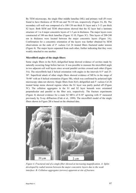
Confirmation for a concentric orientation <strong>of</strong> the layers was further obtained by SEM<br />
observations on the ends <strong>of</strong> P. radiata Cel 26 treated fibers fractured under tension<br />
(Figure 4). The major layers separated from each other, further indicating that they were<br />
weakly attached to one another.<br />
Micr<strong>of</strong>ibril angles <strong>of</strong> the single fibers<br />
Some single fibers in the H2O2 delignified <strong>hemp</strong> showed evidence <strong>of</strong> cavities made by<br />
naturally occurring fungi before harvest. It was possible to measure the micr<strong>of</strong>ibril angle<br />
in two adjacent cell wall layers since several parallel cavities crossed each other (Figure<br />
5A). The micr<strong>of</strong>ibrils had Z helical orientation with angles in the intervals 0-5° and 25-<br />
30°. Superficial attack <strong>of</strong> other single fibers showed evidence <strong>of</strong> MFAs in the range <strong>of</strong><br />
70-90° with an S helical orientation (Figure 5B), which was confirmed by polarized light<br />
microscopy (data not shown). Single fibers from H2O2 delignified and P. radiata Cel 26<br />
treated <strong>hemp</strong> stems showed regions where the S1 layer was partly peeled <strong>of</strong>f (Figure<br />
5C). The cellulose aggregates in the S1 and S2 layer beneath were orientated<br />
perpendicular and parallel to the fiber axis, respectively. The fracture experiments<br />
(Figure 4) showed evidence for a main S2 MFA <strong>of</strong> 0-10° agreeing with 4° measured<br />
previously by X-ray diffraction (Fink et al., 1999). The micr<strong>of</strong>ibril model <strong>of</strong> the single<br />
fibers shown in Figure 2B is based on the obtained data.<br />
Figure 4. Fractured end <strong>of</strong> a single fiber showed at increasing magnification. A: Splits<br />
developed by radial tension between the major concentric layers due to the weak<br />
interface. B: Cellulose aggregates (arrows) apparent at site <strong>of</strong> fracture.<br />
Risø-PhD-11 97