NUI Galway – UL Alliance First Annual ENGINEERING AND - ARAN ...
NUI Galway – UL Alliance First Annual ENGINEERING AND - ARAN ...
NUI Galway – UL Alliance First Annual ENGINEERING AND - ARAN ...

Create successful ePaper yourself
Turn your PDF publications into a flip-book with our unique Google optimized e-Paper software.
An examination into how stem cells form bone in vivo<br />
Birmingham E. 1 , Niebur G. 2 , McHugh P.E. 1 , McNamara L. 1<br />
1 Department of Mechanical and Biomedical Engineering, and National Centre for Biomedical Engineering<br />
Science, <strong>NUI</strong> <strong>Galway</strong>, Ireland. e.birmingham1@nuigalway.ie<br />
2 Department of Aerospace and Mechanical Engineering, University of Notre Dame, Notre Dame, IN<br />
46556, USA.<br />
Abstract<br />
Stem cells are capable of forming different types of<br />
tissues such as bone, cartilage and fat when they<br />
receive appropriate cues, such as biochemicals and<br />
mechanical loading. However, the precise cues that<br />
control the tissues that stem cells ultimately grow<br />
remain unclear. The aim of this study is to examine<br />
what are the natural signals stem cells receive from<br />
their environment which stimulate them to form bone.<br />
1. Introduction.<br />
Mesenchymal stem cells (MSCs) within the marrow<br />
of bone are subjected to a unique microenvironment<br />
known as the stem cell niche. Here MSCs can<br />
experience not only biochemical signalling from<br />
surrounding support cells (osteocytes, osteoblasts,<br />
fibroblasts, adipocytes etc.) but also a mechanical<br />
loading influenced by the trabecular bone structure and<br />
marrow composition (1). While both biochemical<br />
signalling from support cells and mechanical loading<br />
have been shown to direct stem cell differentiation in<br />
vitro, their influence within the niche remains largely<br />
unknown.<br />
Our objectives are (a) to examine the role of support<br />
cells on the differentiation of MSCs and (b) to develop<br />
an in vitro model which allows us to apply<br />
physiological loads to MSCs as they reside in the niche.<br />
2. Materials and Methods.<br />
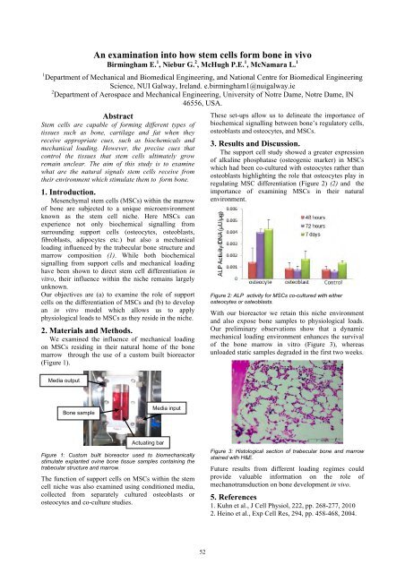
We examined the influence of mechanical loading<br />
on MSCs residing in their natural home of the bone<br />
marrow through the use of a custom built bioreactor<br />
(Figure 1).<br />
Media output<br />
Bone sample<br />
Actuating bar<br />
Media input<br />
Figure 1: Custom built bioreactor used to biomechanically<br />
stimulate explanted ovine bone tissue samples containing the<br />
trabecular structure and marrow.<br />
The function of support cells on MSCs within the stem<br />
cell niche was also examined using conditioned media,<br />
collected from separately cultured osteoblasts or<br />
osteocytes and co-culture studies.<br />
52<br />
These set-ups allow us to delineate the importance of<br />
biochemical signalling between bone’s regulatory cells,<br />
osteoblasts and osteocytes, and MSCs.<br />
3. Results and Discussion.<br />
The support cell study showed a greater expression<br />
of alkaline phosphatase (osteogenic marker) in MSCs<br />
which had been co-cultured with osteocytes rather than<br />
osteoblasts highlighting the role that osteocytes play in<br />
regulating MSC differentiation (Figure 2) (2) and the<br />
importance of examining MSCs in their natural<br />
environment.<br />
Figure 2: ALP activity for MSCs co-cultured with either<br />
osteocytes or osteoblasts.<br />
With our bioreactor we retain this niche environment<br />
and also expose bone samples to physiological loads.<br />
Our preliminary observations show that a dynamic<br />
mechanical loading environment enhances the survival<br />
of the bone marrow in vitro (Figure 3), whereas<br />
unloaded static samples degraded in the first two weeks.<br />
Figure 3: Histological section of trabecular bone and marrow<br />
stained with H&E.<br />
Future results from different loading regimes could<br />
provide valuable information on the role of<br />
mechanotransduction on bone development in vivo.<br />
5. References<br />
1. Kuhn et al., J Cell Physiol, 222, pp. 268-277, 2010<br />
2. Heino et al., Exp Cell Res, 294, pp. 458-468, 2004.