NUI Galway – UL Alliance First Annual ENGINEERING AND - ARAN ...
NUI Galway – UL Alliance First Annual ENGINEERING AND - ARAN ...
NUI Galway – UL Alliance First Annual ENGINEERING AND - ARAN ...
You also want an ePaper? Increase the reach of your titles
YUMPU automatically turns print PDFs into web optimized ePapers that Google loves.
Multi-Scale Computational Modelling of<br />
Physiological Loading on Bone Cells<br />
Verbruggen, S., McNamara, L.M.<br />
Department of Mechanical & Biomedical Engineering, <strong>NUI</strong>, <strong>Galway</strong>, Ireland<br />
email: s.verbruggen1@nuigalway.ie<br />
Abstract<br />
Bone is an adaptive material that can alter its structure<br />
in response to changes in the mechanical environment.<br />
It is possible to enhance bone regeneration in vitro by<br />
manipulating the mechanical environment. However the<br />
mechanical environment necessary to regulate bone<br />
growth is not fully understood. The objective of this<br />
research is to use computational methods to predict the<br />
mechanical micro-environment of bone cells in healthy,<br />
structured bone and thereby inform tissue regeneration<br />
approaches that can mimic the native mechanical<br />
environment to optimise bone regeneration in vitro..<br />
1. Introduction<br />
Bone is an adaptive material, which is particularly<br />
responsive to mechanical loading. [1] Mechanobiology<br />
refers to the ability of biological cells to sense and<br />
respond to extracellular mechanical stimuli. It is<br />
believed that the pathogenesis of diseases such as<br />
osteoporosis, arthritis and diabetes may be correlated to<br />
the mechanobiology of bone cells. [2] Additionally it has<br />
been demonstrated that it is possible to manipulate<br />
cellular responses using mechanical loading regimes,<br />
with such advances paving the way for developing<br />
clinical treatments to address many diseases. [3] However<br />
the precise mechanical environment that regulates<br />
normal bone growth in the body are not fully<br />
understood, because experimental studies to quantify<br />
this are unfeasible.<br />
2. Materials and Methods<br />
Computational models were generated using<br />
ABAQUS® finite element software, with three<br />
complimentary levels of analysis of stimuli in vivo:<br />
Organ-Level: The geometry was generated from CT<br />
(256µm resolution) scans of a femur bone, with material<br />
properties obtained from literature (E=17GPa,<br />
ν=0.38). [3] Loading conditions were applied at the<br />
femoral head, imitating forces generated during normal<br />
walking. The bone was constrained at the distal end.<br />
Tissue-Level: A trabecular and cortical bone tissue<br />
model was generated from µCT (5µm resolution) scans<br />
of a tibia bone using a custom-designed voxel-meshing<br />
software (FEEBE) [4] developed at <strong>NUI</strong>G. The material<br />
properties and mechanical loading was defined from the<br />
organ-level model to simulate stress during normal<br />
walking.<br />
Cellular-Level: An idealised geometry of the lacunarcanalicular<br />
system was generated, with mechanical<br />
loading defined from the tissue-level to represent stress<br />
and strain in the bone matrix surrounding the bone cell.<br />
A model of the osteocyte cell membrane (E=100kPa,<br />
67<br />
ν=0.5) [5] and the surrounding interstitial matrix has also<br />
been developed, allowing the mechanical stimulation of<br />
the cell by its environment to be quantified.<br />
3. Preliminary Results<br />
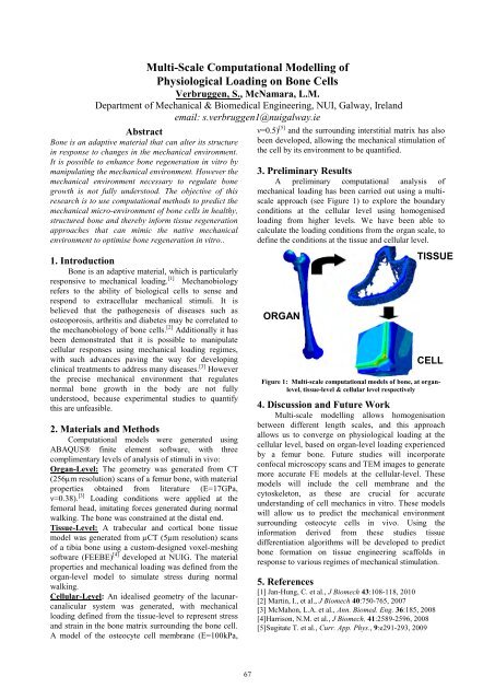
A preliminary computational analysis of<br />
mechanical loading has been carried out using a multiscale<br />
approach (see Figure 1) to explore the boundary<br />
conditions at the cellular level using homogenised<br />
loading from higher levels. We have been able to<br />
calculate the loading conditions from the organ scale, to<br />
define the conditions at the tissue and cellular level.<br />
ORGAN<br />
TISSUE<br />
CELL<br />
Figure 1: Multi-scale computational models of bone, at organlevel,<br />
tissue-level & cellular level respectively<br />
4. Discussion and Future Work<br />
Multi-scale modelling allows homogenisation<br />
between different length scales, and this approach<br />
allows us to converge on physiological loading at the<br />
cellular level, based on organ-level loading experienced<br />
by a femur bone. Future studies will incorporate<br />
confocal microscopy scans and TEM images to generate<br />
more accurate FE models at the cellular-level. These<br />
models will include the cell membrane and the<br />
cytoskeleton, as these are crucial for accurate<br />
understanding of cell mechanics in vitro. These models<br />
will allow us to predict the mechanical environment<br />
surrounding osteocyte cells in vivo. Using the<br />
information derived from these studies tissue<br />
differentiation algorithms will be developed to predict<br />
bone formation on tissue engineering scaffolds in<br />
response to various regimes of mechanical stimulation.<br />
5. References<br />
[1] Jan-Hung, C. et al., J Biomech 43:108-118, 2010<br />
[2] Martin, I., et al., J Biomech 40:750-765, 2007<br />
[3] McMahon, L.A. et al., Ann. Biomed. Eng. 36:185, 2008<br />
[4]Harrison, N.M. et al., J Biomech, 41:2589-2596, 2008<br />
[5]Sugitate T. et al., Curr. App. Phys., 9:e291-293, 2009


