NUI Galway – UL Alliance First Annual ENGINEERING AND - ARAN ...
NUI Galway – UL Alliance First Annual ENGINEERING AND - ARAN ...
NUI Galway – UL Alliance First Annual ENGINEERING AND - ARAN ...

You also want an ePaper? Increase the reach of your titles
YUMPU automatically turns print PDFs into web optimized ePapers that Google loves.
Development of a Micropipette Aspiration and Microscopy System to<br />
Investigate Active Cytoskeletal Remodelling<br />
Reynolds, N. 1 , McGarry J.P. 1<br />
1 Department of Mechanical and Biomedical Engineering, National University of Ireland, <strong>Galway</strong><br />
n.reynolds2@nuigalway.ie<br />
Abstract<br />
Remodelling of the active cytoskeleton plays a critical<br />
role in the response of cells to mechanical stimuli.<br />
Furthermore, the mechanical environment plays an<br />
important role in cell differentiation [1]. Previous<br />
studies have investigated the response of cells to<br />
micropipette aspiration [2-4]. However, the active<br />
response of the actin cytoskeleton has not been<br />
considered. This study will investigate changes in the<br />
actin cytoskeleton during micropipette aspiration of<br />
spread and round cells. The experimental results will be<br />
used to guide the development of an active formulation<br />
of cytoskeletal remodelling in response to external<br />
loading [5]. Detailed examination of the actin<br />
cytoskeleton will also provide insight into remodelling<br />
mechanisms.<br />
1. Introduction<br />
In order to elucidate the key biochemical processes<br />
underlying the experimentally observed phenomena, it<br />
is necessary to characterise dynamic changes in the<br />
cytoskeleton. Micropipette aspiration in tandem with a<br />
novel imaging technique will examine the evolving cell<br />
microstructure under mechanical stimuli.<br />
2. Materials and Methods<br />
Computational: A finite element parametric study of<br />
micropipette aspiration of viscoelastic cells was<br />
performed. The effect of micropipette diameter, nucleus<br />
diameter and vacuum pressure on cell aspiration length<br />
was investigated.<br />
Experimental: Cells will be aspirated with<br />
micropipettes of 5, 10 and 20 µm internal diameter<br />
attached to a custom built pressure control system. The<br />
vacuum pressure applied to the micropipette is<br />
generated from the pressure head difference between<br />
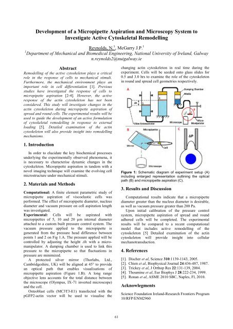
points 1 and 2 on Fig 1.A. The pressure applied will be<br />
controlled by adjusting the height Δh with a micromanipulator.<br />
A damping chamber is used to link this<br />
pressure to the micropipette so that fluctuations in<br />
pressure are minimized.<br />
A protected silver mirror (Thorlabs, Ltd.,<br />
Cambridgeshire, UK) will be aligned at 45° to provide<br />
an optical path that enables visualisations of<br />
micropipette aspiration (Figure 1.B). A long range<br />
objective lens accounts for the total distance between<br />
the microscope (Olympus, IX-71 inverted microscope)<br />
and the cell.<br />
Osteoblast cells (MC3T3-E1) transfected with the<br />
pGFP2-actin vector will be used to visualise the<br />
61<br />
changing actin cytoskeleton in real time during the<br />
experiment. Cells will be seeded onto glass slides for<br />
0.5 and 3.0 hrs to examine the role of the cytoskeleton<br />
in round and spread cell geometries respectively.<br />
Figure 1: Schematic diagram of experiment setup (A)<br />
including enlarged representation outlining the optical<br />
path (B) and micropipette aspiration (C).<br />
3. Results and Discussion<br />
Computational results indicate that a micropipette<br />
diameter greater than the nucleus diameter is desirable,<br />
as well as vacuum pressure greater than 200 Pa.<br />
Upon initial calibration of the pressure control<br />
system, micropipette aspiration of spread and round<br />
adhered cells will be completed. The experimental<br />
results will be compared to a recent computational<br />
model that includes active remodelling of the<br />
cytoskeleton [5] Detailed examination of the actin<br />
cytoskeleton will provide insight into cellular<br />
mechanotransduction.<br />
4. References<br />
[1]. Discher et al, Science 310:1139-1143, 2005.<br />
[2]. Chien et al, Biophysical Journal 24:436-487, 1987.<br />
[3]. Trickey et al, J Orthop Res 22:131-139, 2004.<br />
[4]. Thoumine et al, Eur Biophys J 28:222-234, 1999.<br />
[5]. Ronan et al, ASME 2010 SBC, Naples, Fl, 2010.<br />
Acknowlegments<br />
Science Foundation Ireland-Research Frontiers Program<br />
10/RFP/ENM2960