NUI Galway – UL Alliance First Annual ENGINEERING AND - ARAN ...
NUI Galway – UL Alliance First Annual ENGINEERING AND - ARAN ...
NUI Galway – UL Alliance First Annual ENGINEERING AND - ARAN ...

You also want an ePaper? Increase the reach of your titles
YUMPU automatically turns print PDFs into web optimized ePapers that Google loves.
Simulating Actin Cytoskeleton Remodelling in the Shearing of Chondrocytes<br />
Dowling, E.P. 1 , Ronan, W. 1 , Athanasiou, K.A. 2 , McGarry, J.P. 1<br />
1 Department of Mechanical and Biomedical Engineering, <strong>NUI</strong> <strong>Galway</strong><br />
2 Biomedical Engineering Department, University of California, Davis, CA<br />
e.dowling1@nuigalway.ie<br />
Abstract<br />
In this study, the role of actin fibres in the response of<br />
chondrocytes to shear is experimentally determined. An<br />
active model that describes the assembly of the actin<br />
cytoskeleton in response to cell signalling, and the<br />
dissociation of the actin cytoskeleton in response to a<br />
reduction of intracellular tension is used to simulate the<br />
experimental measurements.<br />
1. Introduction<br />
The actin cytoskeleton, formed via the polymerisation<br />
of actin filaments and phosphorylation of myosin motors,<br />
play a crucial role in the response of cells to mechanical<br />
stimuli. In vitro investigations of the response of cells to<br />
mechanical stimuli provide limited insight into these<br />
mechanisms without the use of active computational<br />
modelling.<br />
2. Methods<br />
Experimental: Chondrocytes were isolated from<br />
bovine articular cartilage tissue, seeded onto glass slides<br />
and positioned adjacent to a tungsten probe. A<br />
piezoelectric motor then drove the probe laterally, leading<br />
to deformation of the cell. This shear event was videorecorded<br />
using a CCD camera. Beam theory was used to<br />
determine the reaction force of the cell at various strain<br />
levels. An additional series of shear experiments were<br />
carried out on cells following the disruption of actin fibres<br />
in the cells by the addition of cytochalasin D (2 µM) to the<br />
media.<br />
Computational: The contractile response of the actinmyosin<br />
fibres is captured using a sliding filament model.<br />
The formation of contractile actin-myosin fibres is<br />
parameterised by the activation level η, which is governed<br />
by a first order kinetic equation [1] :<br />
Ck<br />
f ⎡ σ ⎤ k b<br />
η = [ 1−<br />
η]<br />
− ⎢1<br />
− ⎥η<br />
θ ⎣ σ 0 ⎦ θ<br />
This equation describes the assembly of actin-myosin<br />
units in response to a signal C and dissociation due to a<br />
reduction in tension σ. 3D cell geometries are recreated<br />
from in vitro images (Fig.1a).<br />
3. Results<br />
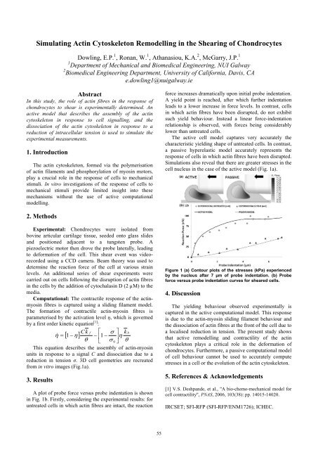
A plot of probe force versus probe indentation is shown<br />
in Fig. 1b. <strong>First</strong>ly, considering the experimental results: for<br />
untreated cells in which actin fibres are intact, the reaction<br />
55<br />
force increases dramatically upon initial probe indentation.<br />
A yield point is reached, after which further indentation<br />
leads to a lower increase in force levels. In contrast, cells<br />
in which actin fibres have been disrupted, do not exhibit<br />
such yield behaviour. Instead a linear force-indentation<br />
relationship is observed, with forces being considerably<br />
lower than untreated cells.<br />
The active cell model captures very accurately the<br />
characteristic yielding shape of untreated cells. In contrast,<br />
a passive hyperelastic model accurately represents the<br />
response of cells in which actin fibres have been disrupted.<br />
Simulations also reveal that there are greater stresses in the<br />
cell nucleus in the case of the active model (Fig. 1a).<br />
Figure 1 (a) Contour plots of the stresses (kPa) experienced<br />
by the nucleus after 7 µm of probe indentation. (b) Probe<br />
force versus probe indentation curves for sheared cells.<br />
4. Discussion<br />
The yielding behaviour observed experimentally is<br />
captured in the active computational model. This response<br />
is due to the actin-myosin sliding filament behaviour and<br />
the dissociation of actin fibres at the front of the cell due to<br />
a localised reduction in tension. The present study shows<br />
that active remodelling and contractility of the actin<br />
cytoskeleton plays a critical role in the deformation of<br />
chondrocytes. Furthermore, a passive computational model<br />
of cell behaviour cannot be used to accurately compute<br />
stresses in a cell or the evolution of the actin cytoskeleton.<br />
5. References & Acknowledgements<br />
[1] V.S. Deshpande, et al., "A bio-chemo-mechanical model for<br />
cell contractility", PNAS, 2006, 103(38): pp. 14015-14020.<br />
IRCSET; SFI-RFP (SFI-RFP/ENM1726); ICHEC.