NUI Galway – UL Alliance First Annual ENGINEERING AND - ARAN ...
NUI Galway – UL Alliance First Annual ENGINEERING AND - ARAN ...
NUI Galway – UL Alliance First Annual ENGINEERING AND - ARAN ...

Create successful ePaper yourself
Turn your PDF publications into a flip-book with our unique Google optimized e-Paper software.
Effects of Knee Flexion on Stented Peripheral Arteries <strong>–</strong> A Computational and In Vitro Study<br />
Ríona Ní Ghriallais 1, 2 1, 2<br />
, Mark Bruzzi<br />
1<br />
Department of Mechanical and Biomedical Engineering, <strong>NUI</strong> <strong>Galway</strong><br />
2<br />
National Centre for Biomedical Engineering Science (NCBES)<br />
r.nighriallais1@nuigalway.ie<br />
Abstract<br />
As stenting in the SFA is not only dependent on stent-artery<br />
contact but also on the relative movement and contact of the<br />
surrounding muscles of the leg, dramatic geometrical changes<br />
of the SFA and PA occur, leading to high failure rates of<br />
stents in these locations. The goal of this work was to<br />
accurately model stent artery-interaction in the SFA by<br />
including the effect of the surrounding muscles during a 90°<br />
knee bend using a computational finite element model.<br />
Furthermore, an in vitro model of the SFA in a system that is<br />
capable of replicating the haemodynamic and biomechanical<br />
environment of the SFA using tissue-engineering principles<br />
was developed. The purpose of the model was to determine<br />
the effect of bending on cell viability, morphology and<br />
orientation in a stented curved vessel.<br />
1. Introduction<br />
The femoropopliteal (FP) artery is a branch of the femoral<br />
artery, the main artery in the upper leg, providing blood to<br />
all muscles and superficial tissues in the thigh. It is the<br />
largest of the femoral artery branches, composed of the<br />
superficial femoral artery (SFA) in the proximal region<br />
and popliteal artery (PA) in the distal region which runs<br />
below the knee. It is characterised by its tortuous<br />
geometry, associating a high atherosclerotic plaque burden<br />
with it. Due to the dynamic forces of the SFA and PA,<br />
peripheral stents are reported to have the highest failure<br />
rates, predominantly due to bending [1]. Worst case<br />
bending can be seen in regions of the SFA/PA behind and<br />
just above the knee [2] and this is detrimental to stent<br />
patency.<br />
2. Methods<br />
3D models of the stented SFA, surroundings muscles<br />
(adductor longus, rectus femoris, biceps femoris, vastus<br />
medialis, vastus laterais and sartorius) and bones (femur<br />
and tibia) were created by importing CT scan data into<br />
MIMICS © software (Materialise NV, Belgium),<br />
constructing anatomically accurate 3D models using each<br />
axial slice. These were then used to create finite element<br />
mesh representations for import into ABAQUS/Explicit.<br />
The stent used for the model was based on the Cordis<br />
SMART Nitinol Stent (OD 7mm, length 20mm). The<br />
analysis involved an initial step where the stent was<br />
deployed in the straight SFA and then a second step where<br />
the knee model (consisting of stented artery, bones,<br />
muscles) was bent to 90°. Medical-grade silicone was used<br />
to produce silicone tubes that were seeded with endothelial<br />
cells to produce pseudovessels. The tubes were coated with<br />
human fibronectin and seeded with HAECs. The<br />
pseudovessels were rotated for 24 h to allow cell adhesion.<br />
Following seeding, a SMART Stent (Cordis) was<br />
deployed in the pseudovessel. The stented model artery<br />
was then transferred into the bioreactor flow loop by<br />
attachment into a specially designed support chamber<br />
which imposed a 50° bend on the tube (the worst case bend<br />
that can be imposed in the tube before kinking occurs).<br />
64<br />
This chamber was then incorporated into the flow loop<br />
which mechanically conditioned the stented model artery<br />
for 24 hours by applying physiological levels of pressure<br />
and flow (120/80 mmHg and 300ml/min respectively) in<br />
an incubator at 37°C [3].<br />
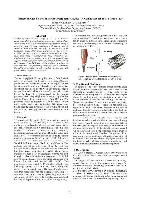
(a)<br />
SFA<br />
(b)<br />
Popliteal<br />
FP Artery<br />
Figure 1. Finite Element Model of Knee Anatomy (a),<br />
femoropopliteal artery (FP) (b) and SMART stent (c)<br />
3. Results and Discussion<br />
The results of the finite element model provide great<br />
insight into the behavior of the artery due to the<br />
surrounding muscles during a knee bend cycle.<br />
Furthermore the incorporation of the stent into the analysis<br />
allows the resulting stress concentrations in the artery due<br />
to the combined in vivo and stent loading to be realized.<br />
Worst case locations of stress in the stented artery after<br />
knee bending can be easily recognized in the distal SFA<br />
region with worst case stress locations of the stented<br />
portion of the artery occurring at the ends of the stent due<br />
to the stiffness mismatch between the ends of the stented<br />
portion and arterial tissue.<br />
In the stented straight control pseudovessel<br />
localized endothelial cell denudation was observed along<br />
the regions where the stent struts were removed. Cells in<br />
between stent strut regions were seen to have altered cell<br />
alignment in comparison to the straight unstented control<br />
which showed all cells in the stimulated model artery to<br />
orient in the longitudinal direction. Comparison of the<br />
response and behaviour in a straight model artery with that<br />
of the cells in the bent model artery highlights the effect of<br />
the bend on cell viability, orientation, morphology and<br />
effects of stiffness mismatch in the stented portion.<br />
4. References<br />
1. D Allie, C Hebert, C Walker, Nitinol stent fractures in<br />
the SFA. Endovascular Today, 2004. July/August Vessel<br />
Update.<br />
2. A Ganguly, A Schneider, B Keck, N Bennett, R Fahirig,<br />
In vivo imaging of superficial femoral artery stents for<br />
deformation analysis. Proceedings of the SPIE 2008, 2008.<br />
6916: p. 69161Y-69161Y-8.<br />
3. M Punchard, ED O'Cearbhaill, JN Mackle, PE McHugh,<br />
TJ Smith, C Stenson-Cox, V Barron Coronary stents:<br />
evaluating the human endothelial cell response in a model<br />
artery in vitro. Annals of Biomedical Engineering, 2009.<br />
(c)