NUI Galway – UL Alliance First Annual ENGINEERING AND - ARAN ...
NUI Galway – UL Alliance First Annual ENGINEERING AND - ARAN ...
NUI Galway – UL Alliance First Annual ENGINEERING AND - ARAN ...

Create successful ePaper yourself
Turn your PDF publications into a flip-book with our unique Google optimized e-Paper software.
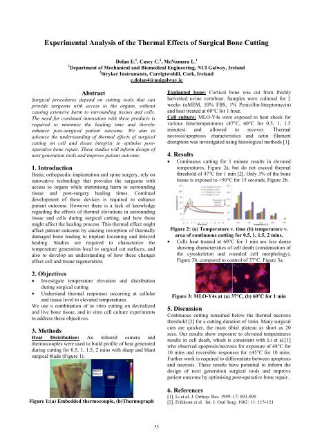
Experimental Analysis of the Thermal Effects of Surgical Bone Cutting<br />
Dolan E. 1 , Casey C. 2 , McNamara L. 1<br />
1 Department of Mechanical and Biomedical Engineering, <strong>NUI</strong> <strong>Galway</strong>, Ireland<br />
2 Stryker Instruments, Carrigtwohill, Cork, Ireland<br />
e.dolan4@nuigalway.ie<br />
Abstract<br />
Surgical procedures depend on cutting tools that can<br />
provide surgeons with access to the organs, without<br />
causing extensive harm to surrounding tissues and cells.<br />
The need for continual innovation with these products is<br />
required to minimise the healing time and thereby<br />
enhance post-surgical patient outcome. We aim to<br />
advance the understanding of thermal effects of surgical<br />
cutting on cell and tissue integrity to optimise postoperative<br />
bone repair. These studies will inform design of<br />
next generation tools and improve patient outcome.<br />
1. Introduction<br />
Brain, orthopaedic implantation and spine surgery, rely on<br />
innovative technology that provides the surgeons with<br />
access to organs while minimising harm to surrounding<br />
tissue and post-surgery healing times. Continual<br />
development of these devices is required to enhance<br />
patient outcome. However there is a lack of knowledge<br />
regarding the effects of thermal elevations in surrounding<br />
tissue and cells during surgical cutting, and how these<br />
might affect the healing process. This thermal effect might<br />
affect patient outcome by causing resorption of thermally<br />
damaged bone leading to implant loosening and delayed<br />
healing. Studies are required to characterize the<br />
temperature generation local to surgical cut surfaces, and<br />
also to develop an understanding of how these changes<br />
effect cell and tissue regeneration.<br />
2. Objectives<br />
Investigate temperature elevation and distribution<br />
during surgical cutting<br />
Understand thermal responses occurring at cellular<br />
and tissue level to elevated temperatures.<br />
We use a combination of in vitro cutting on devitalized<br />
and live bone tissue, and in vitro cell culture experiments<br />
to address these objectives.<br />
3. Methods<br />
Heat Distribution: An infrared camera and<br />
thermocouples were used to build profile of heat generated<br />
during cutting for 0.5, 1, 1.5, 2 mins with sharp and blunt<br />
surgical blade (Figure 1).<br />
Figure 1:(a) Embedded thermocouple, (b)Thermograph<br />
53<br />
Explanted bone: Cortical bone was cut from freshly<br />
harvested ovine vertebrae. Samples were cultured for 2<br />
weeks (αMEM, 10% FBS, 1% Penicillin-Streptomycin)<br />
and heat treated at 60°C for 1 hour.<br />
Cell culture: MLO-Y4s were exposed to heat shock for<br />
various time/temperatures (47°C, 60°C for 0.5, 1, 1.5<br />
minutes) and allowed to recover. Thermal<br />
necrosis/apoptosis characteristics and actin filament<br />
disruption was investigated using histological methods [1].<br />
4. Results<br />
Continuous cutting for 1 minute results in elevated<br />
temperatures, Figure 2a, but do not exceed thermal<br />
threshold of 47°C for 1 min [2]. Only 3% of the bone<br />
tissue is exposed to >50°C for 15 seconds, Figure 2b.<br />
Figure 2: (a) Temperature v. time (b) temperature v.<br />
area of continuous cutting for 0.5, 1, 1.5, 2 mins.<br />
Cells heat treated at 60°C for 1 min are less dense<br />
showing characteristics of cell death (condensation of<br />
the cytoskeleton and rounded cell morphology),<br />
Figure 3b, compared to control of 37°C, Figure 3a.<br />
Figure 3: MLO-Y4s at (a) 37°C, (b) 60°C for 1 min<br />
5. Discussion<br />
Continuous cutting remained below the thermal necrosis<br />
threshold [2] for a cutting duration of 1min. Many surgical<br />
cuts are quicker, the main tibial plateau as short as 20<br />
secs. Our results show exposure to elevated temperatures<br />
results in cell death, which is consistent with Li et al.[1]<br />
who observed apoptosis/necrosis for exposure of 48°C for<br />
10 mins and reversible responses for ≤45°C for 10 mins.<br />
Further work is required to differentiate between apoptosis<br />
and necrosis. These results have potential to inform the<br />
design of next generation surgical tools and improve<br />
patient outcome by optimising post-operative bone repair.<br />
6. References<br />
[1]. Li et al. J. Orthop. Res. 1999: 17: 891-899<br />
[2]. Erikkson et al. Int. J. Oral Surg. 1982: 11: 115-121