NUI Galway – UL Alliance First Annual ENGINEERING AND - ARAN ...
NUI Galway – UL Alliance First Annual ENGINEERING AND - ARAN ...
NUI Galway – UL Alliance First Annual ENGINEERING AND - ARAN ...

Create successful ePaper yourself
Turn your PDF publications into a flip-book with our unique Google optimized e-Paper software.
Active Computational Modelling of Cytoskeletal Remodeling During<br />
Compression and Spreading<br />
Ronan, W. 1 , Deshpande, V. S. 2 , McMeeking, R. M. 3 , McGarry, J.P. 1<br />
1 Department of Mechanical and Biomedical Engineering, National University of Ireland, <strong>Galway</strong><br />
2 Department of Engineering, University of Cambridge<br />
3 Department of Mechanical Engineering, University of California, Santa Barbara<br />
email: w.ronan1@nuigalway.ie<br />
Abstract<br />
Cell spreading is governed by two cooperative<br />
cellular processes: the formation of focal adhesions,<br />
and the active remodeling of the actin cytoskeleton as<br />
the cell spreads 1 . The interaction between these two<br />
processes is poorly understood, and previous<br />
computational models have only examined each process<br />
in isolation. We demonstrate that a novel formulation<br />
that captures key biochemical processes can accurately<br />
capture experimentally observed measurements.<br />
1. Introduction<br />
In the present study an active constitutive<br />
formulation for the remodelling and contractile<br />
behaviour of the actin cytoskeleton and focal<br />
adhesions 3,4 is used to simulate cell spreading on a flat<br />
substrate. Additionally this modelling framework is<br />
used to predict the response of round and spread cells to<br />
compression.<br />
2. Materials and methods<br />
The actin-myosin cytoskeleton is formed via the<br />
assembly of myosin and actin filaments into contractile<br />
stress fibre (SF) bundles. This is captured in our<br />
constitutive model by allowing SFs to assemble in any<br />
direction at any point in the cell. The contractile<br />
behavior of SFs due to the cross-bridge cycling of the<br />
actin-myosin pairs is described by a Hill like equation:<br />
The signal induced formation and tension dependent<br />
dissociation of the actin cytoskeleton is captured using a<br />
first order kinetic equation. This equation gives the<br />
dimensionless activation level of a SF bundle, η,<br />
where C is an exponentially decaying signal. This<br />
formulation has been implemented in a finite element<br />
user-defined material. A model that accounts for the<br />
mechano-sensitivity of focal adhesions based on<br />
thermodynamic considerations is coupled with an<br />
exponential cohesive zone model to simulate spreading.<br />
3. Results<br />
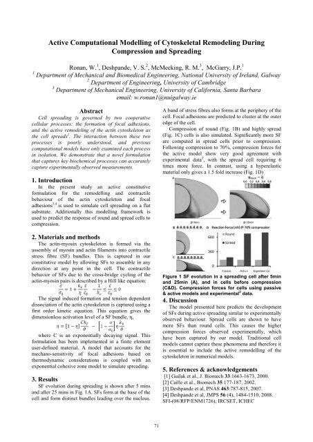
SF evolution during spreading is shown after 5 mins<br />
and after 25 mins in Fig. 1A. SFs form at the base of the<br />
cell and form distinct bundles leading over the nucleus.<br />
71<br />
A band of stress fibres also forms at the periphery of the<br />
cell. Focal adhesions are predicted to cluster at the outer<br />
edge of the cell.<br />
Compression of round (Fig. 1B) and highly spread<br />
(Fig. 1C) cells is also simulated. Significantly more SF<br />
are computed in spread cells prior to compression.<br />
Following compression to 70%, compression forces for<br />
the active model show very good agreement with<br />
experimental data 2 , with the spread cell requiring 6<br />
times more force. In contrast, using a hyperelastic<br />
material only gives a 1.5 fold increase (Fig. 1D)<br />
Figure 1 SF evolution in a spreading cell after 5min<br />
and 25min (A), and in cells before compression<br />
(C&D). Compression forces for cells using passive<br />
& active models and experimental 2 data.<br />
4. Discussion<br />
The model presented here predicts the development<br />
of SFs during active spreading similar to experimentally<br />
observed behaviour. Spread cells are shown to have<br />
more SFs than round cells. This causes the higher<br />
compression forces observed experimentally, which<br />
have been captured by our model. Traditional cell<br />
models cannot capture these phenomena and therefore it<br />
is essential to include the active remodelling of the<br />
cytoskeleton in numerical models.<br />
5. References & acknowledgements<br />
[1] Guilak et al., J. Biomech 33:1663-1673, 2000.<br />
[2] Caille et al., Biomech 35:177-187, 2002.<br />
[3] Deshpande et al, PNAS 463:787-815, 2007.<br />
[4] Deshpande et al, JMPS 56 (4), 1484-1510, 2008.<br />
SFI-(08/RFP/ENM1726), IRCSET, ICHEC