NUI Galway – UL Alliance First Annual ENGINEERING AND - ARAN ...
NUI Galway – UL Alliance First Annual ENGINEERING AND - ARAN ...
NUI Galway – UL Alliance First Annual ENGINEERING AND - ARAN ...
You also want an ePaper? Increase the reach of your titles
YUMPU automatically turns print PDFs into web optimized ePapers that Google loves.
An In vitro Shear Stress System for the Study of Cell-seeded Scaffolds:<br />
A Molecular Biology & Computational Analysis<br />
Meaney, C. L., Piterina A. V., Carroll, G. T., McGloughlin, T. M<br />
Centre for Applied Biomedical Engineering Research (CABER),<br />
Department of Mechanical Aeronautical & Biomedical Engineering,<br />
Materials Surface Science Institute (MSSi), University of Limerick, Ireland<br />
E-mail: claire.meaney@ul.ie<br />
Abstract<br />
Novel materials for vascular graft applications are<br />
continually emerging from research. To attain<br />
comprehensive pre-clinical evaluation, cell-seeded<br />
scaffold materials require exposure to physiological<br />
shear stresses mimetic of those produced in vivo. This<br />
field of testing allows the shear-resistance of the<br />
endothelial layer to be assessed while also providing<br />
indication of anticipated host response to shear through<br />
gene expression analysis. An In vitro methodology was<br />
developed to assess 3D scaffolds in terms of cellular<br />
compatibility under physiological flow conditions. To<br />
ascertain the feasibility of this system computational<br />
and cellular studies were conducted.<br />
1. Introduction<br />
Cellular mechanotransductory pathways are known<br />
to be highly regulated by the mechanical and chemical<br />
properties of the underlying scaffold, thus heightening<br />
the need to assess the cell-material interactions of each<br />
particular material. This highlights the need for an in<br />
vitro platform which facilitates the analysis of 3D<br />
scaffolds under physiological wall shear stress (WSS)<br />
profiles.<br />
2. Bioreactor Characterisation<br />
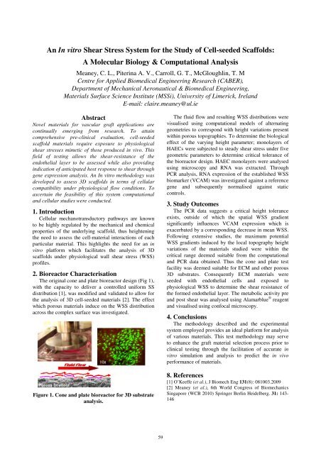
The original cone and plate bioreactor design (Fig 1),<br />
with the capacity to deliver a controlled uniform SS<br />
distribution [1], was modified and validated to allow for<br />
the analysis of 3D cell-seeded materials [2]. The effect<br />
which porous materials induce on the WSS distribution<br />
across the complex surface was investigated.<br />
Figure 1. Cone and plate bioreactor for 3D substrate<br />
analysis.<br />
59<br />
The fluid flow and resulting WSS distributions were<br />
visualised using computational models of alternating<br />
geometries to correspond with height variations present<br />
within porous topographies. To determine the biological<br />
effect of the varying height parameter; monolayers of<br />
HAECs were subjected to steady shear stress under five<br />
geometric parameters to determine critical tolerance of<br />
the bioreactor design. HAEC monolayers were analysed<br />
using microscopy and RNA was extracted. Through<br />
PCR analysis, RNA expression of the established WSS<br />
biomarker (VCAM) was investigated against a reference<br />
gene and subsequently normalised against static<br />
controls.<br />
3. Study Outcomes<br />
The PCR data suggests a critical height tolerance<br />
exists, outside of which the spatial WSS gradient<br />
significantly influences VCAM expression which is<br />
exacerbated by a corresponding decrease in mean WSS.<br />
Following extensive studies, the maximum potential<br />
WSS gradients induced by the local topography height<br />
variations of the materials studied were within the<br />
critical range deemed suitable from the computational<br />
and PCR data obtained. Thus the cone and plate test<br />
facility was deemed suitable for ECM and other porous<br />
3D substrates. Consequently ECM materials were<br />
seeded with endothelial cells and exposed to<br />
physiological WSS to determine the shear resistance of<br />
the formed endothelial layer. The metabolic activity pre<br />
and post shear was analysed using Alamarblue ® reagent<br />
and visualised using confocal microscopy.<br />
4. Conclusions<br />
The methodology described and the experimental<br />
system employed provides an ideal platform for analysis<br />
of various materials. This test methodology may serve<br />
to enhance the graft material selection process prior to<br />
clinical testing through the facilitation of accurate in<br />
vitro simulation and analysis to predict the in vivo<br />
performance of materials.<br />
8. References<br />
[1] O’Keeffe (et al.), J Biomech Eng 131(8): 081003.2009<br />
[2] Meaney (et al.), 6th World Congress of Biomechanics<br />
Singapore (WCB 2010) Springer Berlin Heidelberg. 31: 143-<br />
146


