April - June 2007 - Kasetsart University
April - June 2007 - Kasetsart University
April - June 2007 - Kasetsart University

Create successful ePaper yourself
Turn your PDF publications into a flip-book with our unique Google optimized e-Paper software.
was collected and washed with 70% ethyl alcohol.<br />
After centrifuging at 10,000 rpm for 10 min, the<br />
pellet was dried and suspended in 20-30 µl of<br />
sterile distilled water.<br />
The purified target fragment from the<br />
previous experiment was labeled with<br />
digoxigenin-11-dUTP (Dig-11-dUTP) by using<br />
10xDIG-11-dUTP mixs. The procedure was as<br />
follows: the DNA template was diluted to 50 ng<br />
and prepared for the 50 µl labeling reaction<br />
containing 1µl of DNA template, 5µl of 10x PCR<br />
buffer (200mM TrisHCl, 500mM KCl, 20mM<br />
MgCl 2), 5µl of 10x PCR DIG labeling, 2µl of<br />
each 20 pmole/µl primer and 1µl of Taq DNA<br />
Polymerase (5 units/µl). The labeling PCR<br />
product was separated as described above. The<br />
labeled DNA probe was stored at -20°C and<br />
denatured by heating in boiling water for 10 min<br />
and immediately chilled on ice for 5 min before<br />
use.<br />
The agarose gel containing PCR products<br />
was depurinated in 0.25% HCl for 30 min and<br />
neutralized in 0.4M NaOH for 15 min, and<br />
transferred to Highbond N + nylon membrane by<br />
alkaline 0.4N NaOH. DNAs were fixed under UV<br />
transilluminator for 2.5 min to crosslink the DNA<br />
to the membrane, and washed as suggested by its<br />
manufacturer (Roche ® ). The membrane was<br />
placed into a hybridization bottle containing 3 ml<br />
hybridization solution containing 1% blocking<br />
solution. After incubating for 1 hr at 65°C the<br />
hybridization solution was replaced with a new<br />
hybridization solution containing labeled DNA<br />
probe and incubated at 65°C for additional 18-24<br />
hr. The membrane was removed and washed on a<br />
rotary shaker in solution I (2xSSC, 0.1% SDS) at<br />
65°C for 5 min. The solution was replaced and<br />
the membrane was washed for additional 15 min.<br />
Finally the membrane was washed twice<br />
consecutively with solution II (1xSSC, 0.1%SDS)<br />
and solution III (0.5xSSC, 0.1%SDS) for 15 min<br />
each at 65°C.<br />
<strong>Kasetsart</strong> J. (Nat. Sci.) 41(2) 267<br />
After being washed briefly in washing<br />
buffer and 30 min in 1% blocking buffer, the<br />
membrane was transferred to anti-digoxigenin<br />
alkaline phosphatase conjugated (Roche ® ) and<br />
incubated on a rotary shaker for 45 min at room<br />
temperature. The membrane was washed two<br />
times with washing buffer for 15 min before being<br />
transferred to plastic bag. After adding 500 µl<br />
CDP-Star solution, the bag was sealed, placed into<br />
a Kodak ® x-ray cassette and moved to the dark<br />
room for the detection step. X-ray film was cut to<br />
the proper size and placed over of the membrane<br />
and the closed cassette for 30-60 sec. The film<br />
was transferred to the developer solution until the<br />
band was visible. After washing briefly in water,<br />
the film was removed to the fixer solution until<br />
the background was clear. Finally the film was<br />
washed briefly in water and dried at room<br />
temperature before being photographed.<br />
RESULTS<br />
Bacterial strains<br />
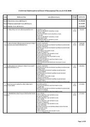
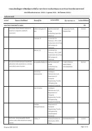
X. citri subsp. citri strains of Thailand<br />
were isolated from different kinds of Citrus spp.,<br />
namely, mandarin, (C. reticulata), lime (C.<br />
aurantifolia), pummelo (C. grandis) and sweet<br />
orange (C. sinensis) from major citrus producing<br />
provinces of Thailand (Table 1). Total X. citri<br />
subsp. citri strains in this study were 19 strains<br />
from Thailand, 2 strains from Japan, and 2 strains<br />
from Saudi Arabia. Other xanthomonads included<br />
in this study consisted of 10 strains of X. fuscans<br />
subsp. aurantifolii, 2 strains of X. alfalfae subsp.<br />
citrumelonis, 1 strain of X. campestris pv.<br />
campestris, 11 strains of X. campestris pv. glycines,<br />
9 strains of X. citri subsp. malvacearum and 1<br />
strain of X. fuscans subsp. fuscans.<br />
PCR specificity<br />
The specific 354-bp PCR fragment was<br />
amplified with 354 F/R primers from all 23 strains<br />
of Xsc (Table 2 and Figure 1A). No fragment of