Workshop proceeding - final.pdf - Faculty of Information and ...
Workshop proceeding - final.pdf - Faculty of Information and ...
Workshop proceeding - final.pdf - Faculty of Information and ...

Create successful ePaper yourself
Turn your PDF publications into a flip-book with our unique Google optimized e-Paper software.
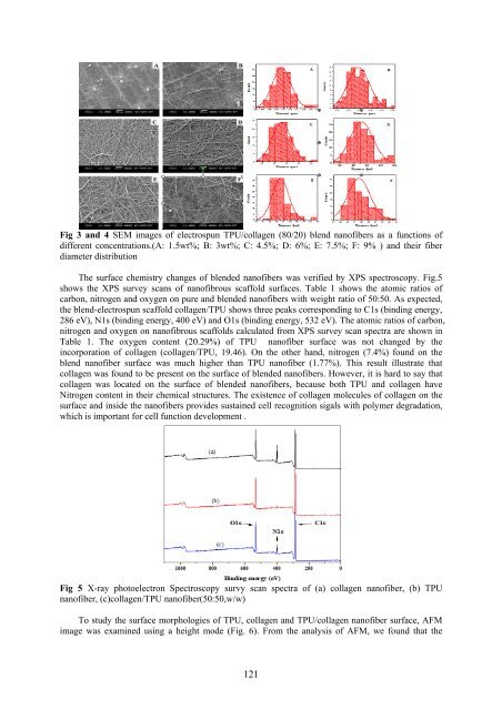
Fig 3 <strong>and</strong> 4 SEM images <strong>of</strong> electrospun TPU/collagen (80/20) blend nan<strong>of</strong>ibers as a functions <strong>of</strong><br />
different concentrations.(A: 1.5wt%; B: 3wt%; C: 4.5%; D: 6%; E: 7.5%; F: 9% ) <strong>and</strong> their fiber<br />
diameter distribution<br />
The surface chemistry changes <strong>of</strong> blended nan<strong>of</strong>ibers was verified by XPS spectroscopy. Fig.5<br />
shows the XPS survey scans <strong>of</strong> nan<strong>of</strong>ibrous scaffold surfaces. Table 1 shows the atomic ratios <strong>of</strong><br />
carbon, nitrogen <strong>and</strong> oxygen on pure <strong>and</strong> blended nan<strong>of</strong>ibers with weight ratio <strong>of</strong> 50:50. As expected,<br />
the blend-electrospun scaffold collagen/TPU shows three peaks corresponding to C1s (binding energy,<br />
286 eV), N1s (binding energy, 400 eV) <strong>and</strong> O1s (binding energy, 532 eV). The atomic ratios <strong>of</strong> carbon,<br />
nitrogen <strong>and</strong> oxygen on nan<strong>of</strong>ibrous scaffolds calculated from XPS survey scan spectra are shown in<br />
Table 1. The oxygen content (20.29%) <strong>of</strong> TPU nan<strong>of</strong>iber surface was not changed by the<br />
incorporation <strong>of</strong> collagen (collagen/TPU, 19.46). On the other h<strong>and</strong>, nitrogen (7.4%) found on the<br />
blend nan<strong>of</strong>iber surface was much higher than TPU nan<strong>of</strong>iber (1.77%). This result illustrate that<br />
collagen was found to be present on the surface <strong>of</strong> blended nan<strong>of</strong>ibers. However, it is hard to say that<br />
collagen was located on the surface <strong>of</strong> blended nan<strong>of</strong>ibers, because both TPU <strong>and</strong> collagen have<br />
Nitrogen content in their chemical structures. The existence <strong>of</strong> collagen molecules <strong>of</strong> collagen on the<br />
surface <strong>and</strong> inside the nan<strong>of</strong>ibers provides sustained cell recognition sigals with polymer degradation,<br />
which is important for cell function development .<br />
Fig 5 X-ray photoelectron Spectroscopy survy scan spectra <strong>of</strong> (a) collagen nan<strong>of</strong>iber, (b) TPU<br />
nan<strong>of</strong>iber, (c)collagen/TPU nan<strong>of</strong>iber(50:50,w/w)<br />
To study the surface morphologies <strong>of</strong> TPU, collagen <strong>and</strong> TPU/collagen nan<strong>of</strong>iber surface, AFM<br />
image was examined using a height mode (Fig. 6). From the analysis <strong>of</strong> AFM, we found that the<br />
121