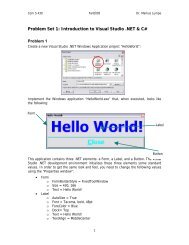
Workshop proceeding - final.pdf - Faculty of Information and ...
Workshop proceeding - final.pdf - Faculty of Information and ...
Workshop proceeding - final.pdf - Faculty of Information and ...

Create successful ePaper yourself
Turn your PDF publications into a flip-book with our unique Google optimized e-Paper software.
2.1 Materials<br />
Sprague-Dauley(SD) male rats,(250-300g, 2-3 month old) were provided from experimental<br />
animal center <strong>of</strong> Sichuan university.(Sichuan, China). Human recombinant mini-TyrRS <strong>and</strong> mini-<br />
TrpRS were from aTyr Pharma (La Jolla, CA).Ⅷ factor related antigen <strong>and</strong> antibody was from<br />
Boster(Wuhan, china). Immunohistochemisty staining kit was purchased from Zhongshan<br />
Goldenbridge (Beijing, China). Revert Aid First Str<strong>and</strong> cDNA Synthesis Kit was purchased from<br />
MBI Company (Lithuania).<br />
2.2.Ethics<br />
All animal procedures were conducted with prior institutional ethical approval under the<br />
requirements <strong>of</strong> the Chinese Prevention <strong>of</strong> Cruelty to Animals Act <strong>and</strong> the Code <strong>of</strong> Practice for the<br />
Care <strong>and</strong> Use <strong>of</strong> Animals for Scientific Purposes. Prior clearance was obtained from the Animal<br />
Experimentation Ethics Committees <strong>of</strong> West china Medical Centre <strong>and</strong> Institutes <strong>of</strong> Animal Science.<br />
The animals <strong>of</strong> this study were inspected by members <strong>of</strong> the West china Medical Centre Animal Ethics<br />
Committee.<br />
2.3.Left anterior descending (LAD) artery ligation<br />
Animals were anaesthetized with 100g/L chloral hydrate (0.2ml per 100g body weight injected<br />
intraperitoneally).The analgesic buprenorphine was given preoperatively(10-20 µg/kg body<br />
weight).Occlusion <strong>of</strong> the left anterior descending coronary artery(LAD) was performed as previously<br />
reported[11],under sterilec conditions, with minor alterations. Briefly,the anaesthetized animal was<br />
incubated endotracheally in a supine position, <strong>and</strong> ventilated with a Harvard Mouse Mini-<br />
Vent(Harvard apparatus, Marchhugstetten,Germany),which supplied 0.2-0.25ml room air 120 times<br />
per minute. The animal was moved onto its right side, <strong>and</strong> a left thoracotomy in the third intercostal<br />
space was provided access to the beating heart. After removing the pericardium,the LAD was<br />
visualized with a stereomicroscope (Leica MZ6,Heerbrugg,Switzerl<strong>and</strong>),<strong>and</strong> occluded with 8/0<br />
prolene suture. The suture position <strong>of</strong> the LAD coronary artery was 0.3mm distal to the<br />
atrioventricular junction. Occlusion was confirmed by observation <strong>of</strong> left ventricular pallor<br />
immediately post ligation <strong>and</strong> an electrocardiogram was used to observe changes such as widening <strong>of</strong><br />
QRS <strong>and</strong> ST-T segment elevation. The chest was closed, the lungs re-inflate <strong>and</strong> the animal moved to<br />
a prone position until spontaneous breathing occurred. Animal were monitored closely for signs <strong>of</strong><br />
infection at the surgical site; none were observed in any animals.<br />
2.4.Experimental groups<br />
A total <strong>of</strong> 80 rats(200-250g) were divided into four different groups(n=20 per each group, <strong>and</strong><br />
n=5 for each time point).⑴sham group: animals underwent a thoracotomy with removal <strong>of</strong> the<br />
pericardium, but no coronary artery ligation (CAL). No suture was placed in the sham animals’heart,<br />
in order to avoid unintended vessel damage or occlusion; ⑵ CAL group, but no mini-TyrRS/mini-<br />
TrpRS-siRNA injection;⑶: CAL+mini-TyrRS(20μl, twice daily, 600μg. Kg -1 .day -1 ) ⑷: CAL+mini-<br />
TrpRS(20μl, twice daily, 600μg. Kg -1 .day -1 ). Mini-TyrRS or mini-TrpRS were administrated by<br />
coronary artery polyvinyl catheter injection to rats.<br />
2.5.Histologic expression<br />
All rats were killed after ligation 3rd, 7th, 14th, <strong>and</strong> 28th day ,the heart was removed <strong>and</strong> fixed in<br />
fresh 4% paraformaldehyde, pH 7.4.The tissue was processed <strong>and</strong> embedded in paraffin using routine<br />
histological procedures. Five micrometre transverse step sections were collected every 200µm<br />
through the entire ventricle(approximately 10-12 sections per animal),<strong>and</strong> stained with Haematoxylin<br />
<strong>and</strong> Eosin(HE).the cells were observed with an inverted phase-contrast microscope (Olympus, Japan)<br />
<strong>and</strong> photographed. The total tube area were analyzed in 3 different fields at ×400 magnification<br />
2.6.Measurement <strong>of</strong> irreversible ischemic injury<br />
After ligation 3rd, 7th, 14th, <strong>and</strong> 28th day, 1% <strong>of</strong> Evans blue solution (5 mL),was infused into the<br />
abdominal vena cava to delineate the ischemic area at risk <strong>of</strong> the left ventricle. The heart was excised<br />
<strong>and</strong> cross-sectioned from the apex to the atrioventricular groove into four specimens <strong>of</strong> 0.8mm in<br />
thickness with the use <strong>of</strong> a stereoscope. The two middle heart slices were incubated in 2,3,5-<br />
39