Workshop proceeding - final.pdf - Faculty of Information and ...
Workshop proceeding - final.pdf - Faculty of Information and ...
Workshop proceeding - final.pdf - Faculty of Information and ...

Create successful ePaper yourself
Turn your PDF publications into a flip-book with our unique Google optimized e-Paper software.
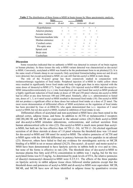
Table 2 The distribution <strong>of</strong> three forms α-MSH in brain tissues by Mass spectrometry analysis<br />
Male<br />
Female<br />
des- mono-act di-act des- mono-act di-act<br />
Hypothalamus + + — + + +<br />
Anterior pituitary + + — — + +<br />
Arcuate nucleus + — — + + —<br />
Neurointermediate lobe + + + + + +<br />
Median eminence + + — + + +<br />
Cerebral cortex + + — + + +<br />
Pre-optic area + + + + + +<br />
Spinal cord — — — — — —<br />
Brain stem — — — — — —<br />
Cerebellum — — — — — —<br />
Discussion<br />
Some researches indicated that no authentic α-MSH was detected in extracts <strong>of</strong> rat brain regions<br />
or human pituitary. In these tissues the only α-MSH variant detected was characterized as des-acetyl<br />
α-MSH. Conversely, acetylated α-MSH was found to be the predominant form in rat pituitary(12). It is<br />
the same result <strong>of</strong> female sheep in our research, Only acetylated forms(including mono-act <strong>and</strong> di-act)<br />
were detected, but except acetylated α-MSH ,we can still find des-acetyl α-MSH in male sheep.<br />
The role <strong>of</strong> the N-acetyl group has been extensively studied in conjunction with<br />
melanocortinergic regulation <strong>of</strong> food intake. Peripheral injection <strong>of</strong> α-MSH in viable yellow obese<br />
mice resulted in significantly lower food intake <strong>and</strong> body weight gain than in mice injected with the<br />
same doses <strong>of</strong> desacetyl-α-MSH (17). Tsujii <strong>and</strong> Bray (16) injected acetyl-α-MSH <strong>and</strong> des-acetyl-α-<br />
MSH intracerebroventricularly (i.c.v.) into food-deprived rats <strong>and</strong> found that acetyl-α-MSH produced<br />
a highly significant reduction <strong>of</strong> food intake at doses <strong>of</strong> 100 <strong>and</strong> 250 pmol whereas des-acetyl-α-MSH<br />
had no effect at any dose between 100 <strong>and</strong> 2500 pmol. Similarly, after i.c.v. administration to fasted<br />
rats, doses <strong>of</strong> 1, 3, or 6 nmol acetyl-α-MSH resulted in decreased food intake (18). Des-acetyl-α-MSH<br />
did not produce a significant effect at these doses but reduced food intake at a dose <strong>of</strong> 25 nmol. The<br />
most recent demonstration <strong>of</strong> differential effects <strong>of</strong> MSH acetylation on the regulation <strong>of</strong> food intake<br />
has been provided by Guo et al.2004(19), who again demonstrated that i.c.v. injection <strong>of</strong> 3 nmol<br />
acetyl-α-MSH, but not des-acetyl-α-MSH, resulted in inhibition <strong>of</strong> food intake in rats.<br />
Differential effects <strong>of</strong> acetylated <strong>and</strong> non-acetylated α-MSH have also been noted in the<br />
adrenal cortex, adipose tissue, <strong>and</strong> bone. In addition to ACTH or melanocortin-2 receptors<br />
(MC2R),MC3R <strong>and</strong> MC5R are expressed in the adrenal cortex (20,21).Both acetyl-α-MSH<br />
<strong>and</strong> des-acetyl-a-MSH stimulate aldosterone, corticosterone, <strong>and</strong> cortisol secretion from<br />
human adrenocortical cells in vitro (22). Des-acetyl-α-MSH is much more potent than acetylα-MSH,<br />
but both are far less effective than ACTH (1–24). ACTH (1–24) elicited significant<br />
secretion <strong>of</strong> all three steroids at doses <strong>of</strong> 1.0 pmol whereas the threshold dose was 1.0 nmol<br />
for des-acetyl-α-MSH <strong>and</strong> 100 nmol for acetyl-α-MSH. The relative potencies <strong>of</strong> ACTH <strong>and</strong><br />
α-MSH agree with the 104-fold difference in potency between the two peptides in rat adrenal<br />
(23). However, others have failed to find any evidence for steroidogenic activity or MC2R<br />
binding <strong>of</strong> α-MSH in rat or mouse adrenal (24,25). Des-acetyl-, di-acetyl- <strong>and</strong> mono-acetyl-α-<br />
MSH have been demonstrated to have lipolytic activity in rabbits both in vivo <strong>and</strong> in vitro,<br />
but none <strong>of</strong> the forms is effective in rats (24). The threshold dose <strong>of</strong> des-acetyl-α-MSH to<br />
increase plasma free fatty acid (FFA) concentration in rabbits was 1 mg/kg, but for the two<br />
acetylated forms was 0.33 mg/kg. As determined by peak levels <strong>of</strong> FFA, the relative potencies<br />
<strong>of</strong> diacetyl-/monoacetyl-/desacetyl-α-MSH were 8.3/5.3/1. The effects <strong>of</strong> the three peptides<br />
on lipolytic activity in rabbit adipose tissue slices followed similar patterns except that the<br />
threshold doses <strong>and</strong> potencies <strong>of</strong> acetyl-α-MSH <strong>and</strong> di-acetyl-α-MSH were equivalent. MC2R,<br />
MC4R, <strong>and</strong> MC5R have now all been identified in adipose tissue by quantitative reverse<br />
58