IGCAR : Annual Report - Indira Gandhi Centre for Atomic Research
IGCAR : Annual Report - Indira Gandhi Centre for Atomic Research
IGCAR : Annual Report - Indira Gandhi Centre for Atomic Research

Create successful ePaper yourself
Turn your PDF publications into a flip-book with our unique Google optimized e-Paper software.
IGC<br />
<strong>Annual</strong> <strong>Report</strong> 2007<br />
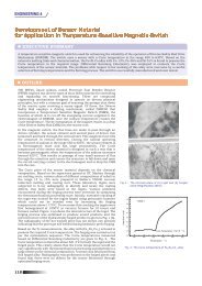
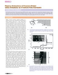
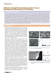
Fig.2 Skull phantom image using Kodak IP (14) and <strong>IGCAR</strong> IP (13)<br />
-<br />
from F I get trapped at bromine<br />
ion vacancies thereby <strong>for</strong>ming<br />
F(Br-) centers. The interstitial<br />
fluorine atoms thus <strong>for</strong>med<br />
combine with the lattice fluorine<br />
-<br />
ions to <strong>for</strong>m molecular ions, F 2<br />
(H type center), whose presence<br />
on x-irradiation was detected by<br />
ESR (Fig. 1b). In the proposed<br />
model, F I-<br />
ions act as hole traps<br />
and bromine vacancies act as<br />
electron centers. On photo<br />
stimulation, the electrons<br />
released from F(Br-) centers<br />
recombine with holes trapped<br />
at F 2 - centers, which results in<br />
intrinsic (self-trapped exciton)<br />
emission (~300 nm) which is<br />
characteristic of the BaFBr host<br />
(Fig. 1c). This radiatively excites<br />
the Eu 2+ ions thereby causing<br />
PSL emission around 400 nm.<br />
The merits of the proposed<br />
mechanism are: it explains why<br />
only fluorine excess compounds<br />
cause intense PSL, it provides a<br />
charge compensation<br />
mechanism arising out of the<br />
presence of bromine vacancies,<br />
it supports the experimentally<br />
-<br />
observed F 2 centers on X-<br />
irradiation in fluorine excess<br />
BaFBr, and above all it brings<br />
the material back to its initial<br />
state on photo stimulation.<br />
An imaging plate has been<br />
made with the help of M/s<br />
Kiran X-ray screens Ltd,<br />
Mumbai using the indigenously<br />
synthesized storage phosphor. A<br />
comparative skull phantom<br />
image was recorded using a<br />
commercial scanner and a<br />
commercial Kodak IP as well as<br />
the IP made from the storage<br />
phosphor synthesized at <strong>IGCAR</strong><br />
at the same X-ray exposure<br />
parameters (Fig. 2). All the<br />
salient features of the image<br />
are seen in the IP made from<br />
our phosphor. The sensitivity is<br />
comparatively good but the<br />
contrast needs improvement.<br />
The grain morphology is<br />
uneven and higher than the<br />
desired value, which causes<br />
uneven light scattering that<br />
reduces the image contrast.<br />
Modified synthesis techniques<br />
and grinding procedure of the<br />
phosphor developed recently<br />
have improved the grain<br />
morphology (grain size ~ 20<br />
µm). Recent ef<strong>for</strong>ts in preparing<br />
the phosphor in large scale with<br />
a simple and cost effective<br />
technique using the local make<br />
chemicals have been<br />
successful. Manufacturing more<br />
number of test IPs and<br />
comparative studies with<br />
commercial IPs are in progress.<br />
BASIC RESEARCH 165