IGCAR : Annual Report - Indira Gandhi Centre for Atomic Research
IGCAR : Annual Report - Indira Gandhi Centre for Atomic Research
IGCAR : Annual Report - Indira Gandhi Centre for Atomic Research

Create successful ePaper yourself
Turn your PDF publications into a flip-book with our unique Google optimized e-Paper software.
IGC<br />
<strong>Annual</strong> <strong>Report</strong> 2007<br />
VI.12. X-ray Diffraction Studies using Imaging Plate as<br />
an Area Detector<br />
Imaging plate based X-ray<br />
area detectors were initially<br />
used <strong>for</strong> protein crystallography<br />
in the 1980s and more recently,<br />
their use has extended to smallmolecule<br />
structural analyses<br />
and powder diffractometry. The<br />
usage of imaging plate is<br />
particularly advantageous <strong>for</strong><br />
the characterization of<br />
polycrystalline materials since it<br />
permits simultaneous collection<br />
of many orders of Bragg<br />
reflections. There is also a<br />
significant size advantage<br />
compared to CCD based area<br />
detector. In addition to the<br />
enormous reduction of data<br />
acquisition time <strong>for</strong> analyses,<br />
two-dimensional diffraction<br />
patterns contain much more<br />
in<strong>for</strong>mation than conventional<br />
linear scans (i.e. - 2 scans)<br />
collected using standard<br />
powder diffractometers. Two<br />
dimensional diffraction patterns<br />
of polycrystalline samples<br />
typically consist of concentric<br />
(Debye-Scherrer) rings<br />
produced by the superposition<br />
of reflections from many<br />
crystals illuminated by the X-ray<br />
beam, which are oriented with<br />
a set of (hkl) crystallographic<br />
planes oriented to fulfill the<br />
Bragg condition. Depending on<br />
sample characteristics, these<br />
rings might be continuous or<br />
spotty and display specific<br />
variation in the intensities along<br />
them. These features contain<br />
important in<strong>for</strong>mation about<br />
the microstructure of the<br />
sample: grain size, preferential<br />
orientation, mosaicity, stress<br />
etc. Additionally, twodimensional<br />
patterns can be<br />
converted into conventional<br />
linear scans by radial or<br />
azimuthal integration of pixel<br />
intensities. The generated linear<br />
scans can be processed as<br />
usual <strong>for</strong> mineral phase<br />
identification, crystallinity or<br />
Rietveld refinement studies.<br />
Nevertheless, during this data<br />
reduction procedure, most of<br />
the in<strong>for</strong>mation regarding the<br />
microstructure of the material is<br />
lost. Hence to get the full<br />
advantage of two-dimensional<br />
diffraction <strong>for</strong> polycrystalline<br />
materials we have to extract the<br />
in<strong>for</strong>mation contained in the<br />
two dimensional diffraction<br />
patterns. Here we are reporting<br />
the diffraction experiment<br />
results obtained using the<br />
imaging plate as an area<br />
detector <strong>for</strong> both single crystal<br />
and powder experiments. The<br />
imaging plate reader has 5<br />
mega pixel resolution <strong>for</strong> 250<br />
mm X 200 mm size plate with<br />
each pixel size of 100 the<br />
data file size is 10 Mbytes.<br />
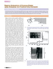
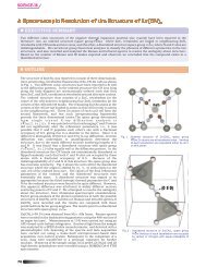
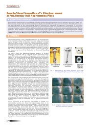
Fig.1 Laue diffraction pattern of a Si single crystal obtained using the<br />
Imaging plate as an area detector (the plate read using reader developed<br />
at MSD <strong>IGCAR</strong>). The inset shows the maximum zoom of a diffraction point<br />
and its intensity profile in surface and line plot.<br />
Laue X-ray diffraction is<br />
historically the first diffraction<br />
method <strong>for</strong> structural<br />
166 BASIC RESEARCH