

iaea human health series publications - SEDIM
iaea human health series publications - SEDIM
iaea human health series publications - SEDIM
- No tags were found...

You also want an ePaper? Increase the reach of your titles
YUMPU automatically turns print PDFs into web optimized ePapers that Google loves.
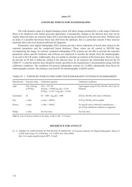
Annex IVEXPOSURE INDICES FOR MAMMOGRAPHYThe wide dynamic range of a digital imaging system will allow images produced by a wide range of detectordoses to be displayed with similar greyscale appearance. Consequently, changes to the detector dose may not bereadily observed unless an exposure index (EI) is provided giving an indication of the previous dose. Without suchan index it is possible that breast doses may drift from the optimum; this is a particular concern if they increasemarkedly above the level deemed sufficient for diagnosis.Fortunately, most digital radiography (DX) systems provide a direct indication of breast dose, based on theexposure parameters and the compressed breast thickness. These values are all carried in DICOM tagsaccompanying the image. In contrast, computed radiography (CR) systems are not able to provide the exposureparameters unless specific hardware and software are employed to transfer the details from the mammographyX ray unit to the CR reader. Additionally, they are unable to calculate an estimate of the breast dose. However, theydo provide an EI that is indirectly related to the detector dose. In all instances the relationship between the EI(Table IV–1) and the detector dose should be clearly specified in the manufacturer’s documentation along with thecalibration conditions. The conditions for general radiographic systems [A–1] differ substantially from those formammography systems. Developing a universal EI for mammography would be useful.TABLE IV–1. EXPOSURE INDICES FOR COMPUTED RADIOGRAPHY SYSTEMS IN MAMMOGRPHYManufacturer Exposure index Calibration equation Calibration conditionsAgfaSAL, SALlogor PVIlogSAL = 253·÷KSALlog = 10 000·log 10 (K) + 8 581PVIlog = 13 287.7·log 10 (K) + 23 824Agfa suggest using 28 kVp, Mo/Mo with 2 mm Alfiltration.Carestream EI EI = 1000 · log 10 (K) + 1000 28 kVp, Mo/Mo with 2 mm Al filtrationFuji s value s value = 2400/K 25 kVp, Mo/Mo with no paddleKonica s value s value = 2400/K No specific advice offered by manufacturer —use of Fuji calibration conditions is suggestedPhilips s value See Fuji entry See Fuji entryNote: K is the air kerma incident on the plate, in mR (1 mR = 8.76 μGy).REFERENCE FOR ANNEX IV[A–1] AMERICAN ASSOCIATION OF PHYSICISTS IN MEDICINE, An Exposure Indicator for Digital Radiography: Report ofAAPM Task Group 116, AAPM Rep. 116, AAPM, New York (2009),http://www.aapm.org/pubs/reports/RPT_116.pdf172

