

iaea human health series publications - SEDIM
iaea human health series publications - SEDIM
iaea human health series publications - SEDIM
- No tags were found...

You also want an ePaper? Increase the reach of your titles
YUMPU automatically turns print PDFs into web optimized ePapers that Google loves.
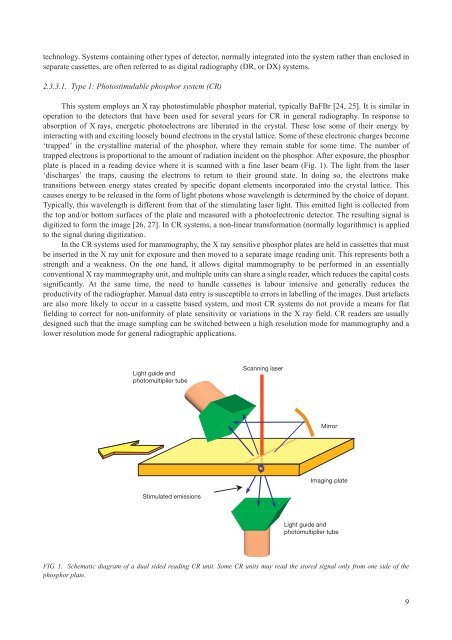
technology. Systems containing other types of detector, normally integrated into the system rather than enclosed inseparate cassettes, are often referred to as digital radiography (DR, or DX) systems.2.3.3.1. Type 1: Photostimulable phosphor system (CR)This system employs an X ray photostimulable phosphor material, typically BaFBr [24, 25]. It is similar inoperation to the detectors that have been used for several years for CR in general radiography. In response toabsorption of X rays, energetic photoelectrons are liberated in the crystal. These lose some of their energy byinteracting with and exciting loosely bound electrons in the crystal lattice. Some of these electronic charges become‘trapped’ in the crystalline material of the phosphor, where they remain stable for some time. The number oftrapped electrons is proportional to the amount of radiation incident on the phosphor. After exposure, the phosphorplate is placed in a reading device where it is scanned with a fine laser beam (Fig. 1). The light from the laser‘discharges’ the traps, causing the electrons to return to their ground state. In doing so, the electrons maketransitions between energy states created by specific dopant elements incorporated into the crystal lattice. Thiscauses energy to be released in the form of light photons whose wavelength is determined by the choice of dopant.Typically, this wavelength is different from that of the stimulating laser light. This emitted light is collected fromthe top and/or bottom surfaces of the plate and measured with a photoelectronic detector. The resulting signal isdigitized to form the image [26, 27]. In CR systems, a non-linear transformation (normally logarithmic) is appliedto the signal during digitization.In the CR systems used for mammography, the X ray sensitive phosphor plates are held in cassettes that mustbe inserted in the X ray unit for exposure and then moved to a separate image reading unit. This represents both astrength and a weakness. On the one hand, it allows digital mammography to be performed in an essentiallyconventional X ray mammography unit, and multiple units can share a single reader, which reduces the capital costssignificantly. At the same time, the need to handle cassettes is labour intensive and generally reduces theproductivity of the radiographer. Manual data entry is susceptible to errors in labelling of the images. Dust artefactsare also more likely to occur in a cassette based system, and most CR systems do not provide a means for flatfielding to correct for non-uniformity of plate sensitivity or variations in the X ray field. CR readers are usuallydesigned such that the image sampling can be switched between a high resolution mode for mammography and alower resolution mode for general radiographic applications.Light guide andphotomultiplier tubeScanning laserMirrorImaging plateStimulated emissionsLight guide andphotomultiplier tubeFIG. 1. Schematic diagram of a dual sided reading CR unit. Some CR units may read the stored signal only from one side of thephosphor plate.9

