

iaea human health series publications - SEDIM
iaea human health series publications - SEDIM
iaea human health series publications - SEDIM
- No tags were found...

Create successful ePaper yourself
Turn your PDF publications into a flip-book with our unique Google optimized e-Paper software.
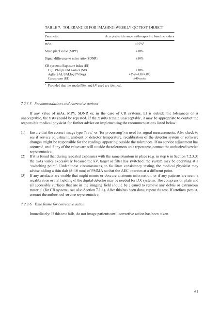
TABLE 7. TOLERANCES FOR IMAGING WEEKLY QC TEST OBJECTParametermAsAcceptable tolerance with respect to baseline values±10% aMean pixel value (MPV) ±10%Signal difference to noise ratio (SDNR) ±10%CR systems: Exposure index (EI)Fuji, Philips and Konica (S#)Agfa (SAL/SALlog/PVIlog)Carestream (EI)±10%±5%/±430/±580±40 unitsaProvided that the anode/filter and kV used are identical.7.2.3.5. Recommendations and corrective actionsIf any value of mAs, MPV, SDNR or, in the case of CR systems, EI is outside the tolerances or isunacceptable, the tests should be repeated. If the results remain unacceptable, it may be appropriate to contact theresponsible medical physicist for further advice on implementing the recommendations listed below:(1) Ensure that the correct image type (‘raw’ or ‘for processing’) is used for signal measurements. Also check tosee if service adjustment, ambient or detector temperature, recalibration of the detector system or softwarechanges might be responsible for the readings appearing outside the tolerances. If no service adjustment hasoccurred, and if any of the values are still outside the tolerances on a repeat test, contact the authorized servicerepresentative.(2) If it is found that during repeated exposures with the same phantom in place (e.g. in step 6 in Section 7.2.3.3)the mAs varies excessively because the kV, target or filter has switched, the system may be operating at a‘switching point’. Under these circumstances, to facilitate consistency testing, the medical physicist mayadvise adding a thin slab (5–10 mm) of PMMA so that the AEC operates at a different point.(3) If any artefacts are visible that might mimic or obscure anatomic information, or if any patterns are seen, arecalibration or flat fielding of the digital detector may be needed for DX systems. The compression plate andall accessible surfaces that are in the imaging field should be cleaned to remove any debris or extraneousmaterial (for CR systems, see also Section 7.1.4). After this has been done, repeat the test. If artefacts persist,contact the authorized service representative.7.2.3.6. Time frame for corrective actionImmediately: If this test fails, do not image patients until corrective action has been taken.61

