You also want an ePaper? Increase the reach of your titles
YUMPU automatically turns print PDFs into web optimized ePapers that Google loves.
DODGE-<strong><strong>LICHEN</strong>S</strong> <strong>AND</strong> <strong>LICHEN</strong> PABABITES I87<br />
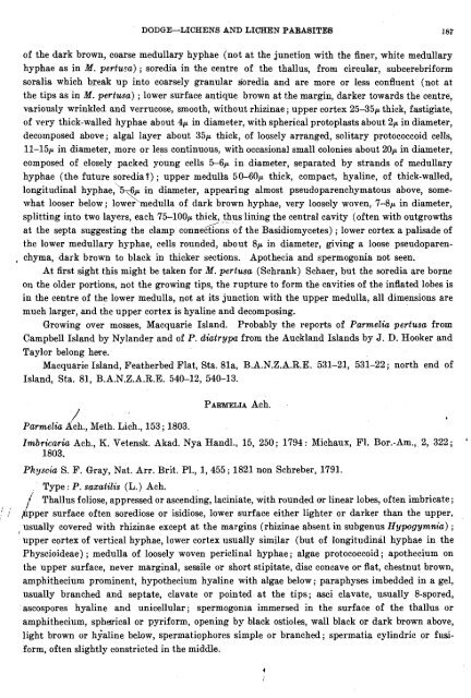
of the dark brown, coarse medullary hyphae (not at the junction with the finer, white medullary<br />
hyphae as in M. pertusa) ; soredia in the centre of the thallus, from circul'ar, subcerebriform<br />
soralia which brealr up into coarsely granular soredia and are more or less confluent (not at<br />
the tips as in M. pertusa) ; lower surface antique brown at the margin, darker towards the centre,<br />
variously wrinkled and verrucose, smooth, without rhizinae ; upper cortex 25-35p thick, fastigiate,<br />
of very thick-walled hyphae about 4p in diameter, with spherical protoplasts about 2p in diameter,<br />
decomposed above; algal layer about 35p thick, of loosely arranged, solitary protococcoid cells,<br />
11-15p in diameter, more or less continuous, with occasional small colonies about 20p in diameter,<br />
composed of closely packed young cells 5-6p in diameter, separated by strands of medullary<br />
hyphae (the future soredia?) ; upper medulla 50-60p thick, compact, hyaline, of thick-walled,<br />
longitudinal hyphae:%-6p in diameter, appearing almost pseudoparenchymatous above, somewhat<br />
looser below; lower \ medulla of dark brown hyphae, very loosely woven, 7-8p in diameter,<br />
splitting into two layers, each 75-100p thick, thus lining the central cavity (often with outgrowths<br />
1.' . -<br />
at the septa suggesting the clamp connections of the Basidiomycetes) ; lower cortex a palisade of<br />
the lower medullary hyphae, cells rounded, about 8p in diameter, giving a loose pseudoparen-<br />
, chyma, ,dark brown to black in thicker sections. Apothecia and spermogonia not seen.<br />
At first sight this might be taken for M. pertusa (Schrank) Schaer, but the soredia are borne<br />
on the older portions, not the growing tips, the rupture to form the cavities of the inflated lobes is<br />
in the centre of the lower medulla, not at its junction with the upper medulla, all dimensions are<br />
much larger, and the upper cortex is hyaline and decomposing.<br />
Growing over mosses, Macquarie Island. Probably the reports of Parmelia pertusa from<br />
Campbell Island by Nylander and of P. diatrypu from the Auckland Islands by J. D. Hooker and<br />
Taylor belong hme.<br />
Macquarie Island, Featherbed Flat, Sta. 81a, B.A.N.Z.A.R.E. 531-21, 531-22; north end of<br />
Island, Sta. 81, B.A.N.Z.A.R.E. 540-12, 540-13.<br />
/'<br />
Parmelia ~ch., Meth. Lich., 153; 1803.<br />
PARMELIA Ach.<br />
Imbricariu Ach., K. Vetenslr. Akad. Nya Handl., 15, 250; 1794: Michaux, F1. Bor.-Am., 2, 322;<br />
1803.<br />
Physcia S. F. Gray, Nat. Arr. Brit. Pl., 1, 455 ; 1821 non Schreber, 1791.<br />
Type : P. sazatilis (L.) Ach.<br />
/<br />
Thallus foliose, appressed or ascending, laciniate, with rounded or linear lobes, often imbricate ;<br />
5 ,<br />
' .i pipper surface often sorediose or isidiose, lower surface either lighter or darker than the upper,<br />
usually covered with rhizinae except at the margins (rhizinae absent in subgenus Hypogyntnia) ;<br />
upper cortex of vertical hyphae, lower cortex usually similar (but of longitudinal hyphae in the<br />
Physcioideae) ; medulla of loosely woven periclinal hyphae ; algae protococcoid ; apothecium on<br />
the upper surface, never marginal, sessile or short stipitate, disc concave or flat, chestnut brown,<br />
amphithecium prominent, hypothecium hyaline with algae below; paraphyses imbedded in a gel,<br />
usually branched and septate, clavate or pointed at the tips; asci clavate, usually 8-spored,<br />
ascospores hyaline and unicellular; spermogonia immersed in the surface of the thallus or<br />
amphithecium, spherical or pyriform, opening by black ostioles, wall black or dark brown above,<br />
light brown or hialine below, spermatiophores simple or branched; spermatia cylindric or fnsiform,<br />
often slightly constricted in the middle.<br />
'


