Retinal Prosthesis Dissertation - Student Home Pages
Retinal Prosthesis Dissertation - Student Home Pages
Retinal Prosthesis Dissertation - Student Home Pages

Create successful ePaper yourself
Turn your PDF publications into a flip-book with our unique Google optimized e-Paper software.
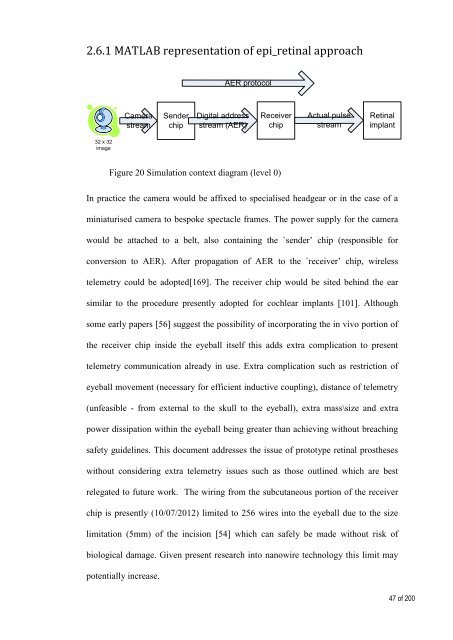
2.6.1 MATLAB representation of epi_retinal approach<br />
AER protocol<br />
Camera<br />
stream<br />
Sender<br />
chip<br />
Digital address<br />
stream (AER)<br />
Receiver<br />
chip<br />
Actual pulse<br />
stream<br />
<strong>Retinal</strong><br />
implant<br />
32 x 32<br />
image<br />
Figure 20 Simulation context diagram (level 0)<br />
In practice the camera would be affixed to specialised headgear or in the case of a<br />
miniaturised camera to bespoke spectacle frames. The power supply for the camera<br />
would be attached to a belt, also containing the `sender’ chip (responsible for<br />
conversion to AER). After propagation of AER to the `receiver’ chip, wireless<br />
telemetry could be adopted[169]. The receiver chip would be sited behind the ear<br />
similar to the procedure presently adopted for cochlear implants [101]. Although<br />
some early papers [56] suggest the possibility of incorporating the in vivo portion of<br />
the receiver chip inside the eyeball itself this adds extra complication to present<br />
telemetry communication already in use. Extra complication such as restriction of<br />
eyeball movement (necessary for efficient inductive coupling), distance of telemetry<br />
(unfeasible - from external to the skull to the eyeball), extra mass\size and extra<br />
power dissipation within the eyeball being greater than achieving without breaching<br />
safety guidelines. This document addresses the issue of prototype retinal prostheses<br />
without considering extra telemetry issues such as those outlined which are best<br />
relegated to future work. The wiring from the subcutaneous portion of the receiver<br />
chip is presently (10/07/2012) limited to 256 wires into the eyeball due to the size<br />
limitation (5mm) of the incision [54] which can safely be made without risk of<br />
biological damage. Given present research into nanowire technology this limit may<br />
potentially increase.<br />
47 of 200