Retinal Prosthesis Dissertation - Student Home Pages
Retinal Prosthesis Dissertation - Student Home Pages
Retinal Prosthesis Dissertation - Student Home Pages

Create successful ePaper yourself
Turn your PDF publications into a flip-book with our unique Google optimized e-Paper software.
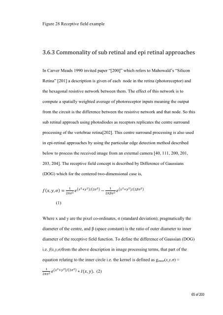
Figure 28 Receptive field example<br />
3.6.3 Commonality of sub retinal and epi retinal approaches<br />
In Carver Meads 1990 invited paper “[200]” which refers to Mahowald’s “Silicon<br />
Retina” [201] a description is given of each node in the retina (photoreceptor) and<br />
the hexagonal resistive network between them. The effect of this network is to<br />
compute a spatially weighted average of photoreceptor inputs meaning the output<br />
from the circuit is the difference between the resistive network and that node. So this<br />
sub retinal approach using photodiodes as receptors replicates the centre surround<br />
processing of the vertebrae retina[202]. This centre surround processing is also used<br />
in epi-retinal approaches by using the particular edge detection method described<br />
below to process the received image from an external camera [40, 111, 200, 201,<br />
203, 204]. The receptive field concept is described by Difference of Gaussians<br />
(DOG) which for the centered two-dimensional case is,<br />
( )<br />
( ) ( ) ( ) ( )<br />
(1)<br />
Where x and y are the pixel co-ordinates, σ (standard deviation); pragmatically the<br />
diameter of the centre, and β (space constant) is the ratio of outer diameter to inner<br />
diameter of the receptive field function. To define the difference of Gaussian (DOG)<br />
i.e. f(x,y,σ)from the above description in image processing terms, that part of the<br />
equation relating to the inner circle i.e. the kernel is defined as g inner (x,y,σ) =<br />
( ) ( )<br />
( ) (2)<br />
65 of 200