Segmentation of Stochastic Images using ... - Jacobs University
Segmentation of Stochastic Images using ... - Jacobs University
Segmentation of Stochastic Images using ... - Jacobs University

You also want an ePaper? Increase the reach of your titles
YUMPU automatically turns print PDFs into web optimized ePapers that Google loves.
Chapter 1<br />
Introduction<br />
The development <strong>of</strong> mathematical methods for image processing became a rapidly growing research<br />
field during the last decades. The fast progress in the speed <strong>of</strong> widely available computer systems<br />
allowed the numerical implementation <strong>of</strong> complex models. A specialty is the development <strong>of</strong> segmentation<br />
algorithms based on partial differential equations (PDEs). The aim <strong>of</strong> a segmentation<br />
algorithm is the decomposition <strong>of</strong> an image into the object and the background. Typically, detecting<br />
edges inside an image or meeting a homogenization criterion for the object and the background lead<br />
to a segmentation. Widely used segmentation approaches are the random walker segmentation [59],<br />
the Mumford-Shah segmentation [107] and the related Ambrosio-Tortorelli regularization [14], and<br />
active contour methods based on level set formulations [30,31,82,138]. Besided these segmentation<br />
methods, which will be investigated in this thesis, there are other segmentation methods like region<br />
growing [127], watersheds [136], snakes [76], and graph cuts [25].<br />
Many applications use segmentation methods, e.g. quality control, machine vision, and medical<br />
image processing. For example, the further treatment for cancer patients bases on the segmented<br />
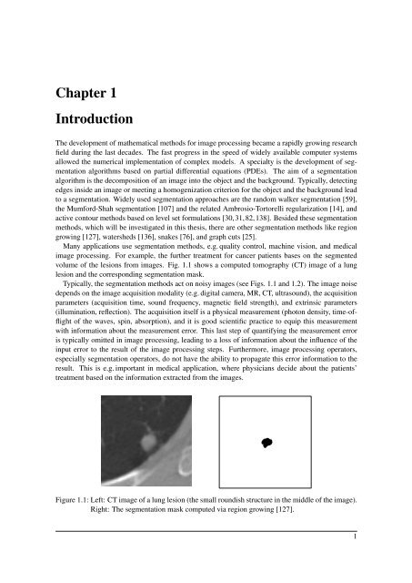
volume <strong>of</strong> the lesions from images. Fig. 1.1 shows a computed tomography (CT) image <strong>of</strong> a lung<br />
lesion and the corresponding segmentation mask.<br />
Typically, the segmentation methods act on noisy images (see Figs. 1.1 and 1.2). The image noise<br />
depends on the image acquisition modality (e.g. digital camera, MR, CT, ultrasound), the acquisition<br />
parameters (acquisition time, sound frequency, magnetic field strength), and extrinsic parameters<br />
(illumination, reflection). The acquisition itself is a physical measurement (photon density, time-<strong>of</strong>flight<br />
<strong>of</strong> the waves, spin, absorption), and it is good scientific practice to equip this measurement<br />
with information about the measurement error. This last step <strong>of</strong> quantifying the measurement error<br />
is typically omitted in image processing, leading to a loss <strong>of</strong> information about the influence <strong>of</strong> the<br />
input error to the result <strong>of</strong> the image processing steps. Furthermore, image processing operators,<br />
especially segmentation operators, do not have the ability to propagate this error information to the<br />
result. This is e.g. important in medical application, where physicians decide about the patients’<br />
treatment based on the information extracted from the images.<br />
Figure 1.1: Left: CT image <strong>of</strong> a lung lesion (the small roundish structure in the middle <strong>of</strong> the image).<br />
Right: The segmentation mask computed via region growing [127].<br />
1