- Page 3 and 4: TABLE OF CONTENTSA. PERIODIC ACTIVI
- Page 5 and 6: 1 PUBLISHABLE EXECUTIVE SUMMARY EUR
- Page 7 and 8: Dr. Davide Gabellini(28) Swiss Inst
- Page 9 and 10: WP7 Molecular characterization of s
- Page 11 and 12: To identify protein-protein interac
- Page 13 and 14: expressed in a particular tissue. T
- Page 15 and 16: Duchenne muscular dystrophy (DMD).
- Page 17 and 18: WP21 Reachout to the broader RNA co
- Page 19 and 20: 4.1 TABLE OF MILESTONES JPA MONTH 2
- Page 21 and 22: Numerous plasmids, constructs, extr
- Page 23 and 24: collaboration between EURASNET and
- Page 25 and 26: 5.1 WORK PACKAGE REPORTSWork Packag
- Page 27 and 28: • Ongoing regular addition of new
- Page 29 and 30: Work Package 4 The Alternative Spli
- Page 31 and 32: Work Package 6 In silico approaches
- Page 33 and 34: alternative splicing.162 Compare sp
- Page 35 and 36: 33 Agreement on pre-mRNA substrates
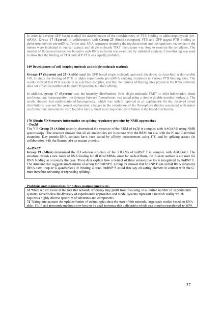
- Page 37: its numerous binding sites on both
- Page 41 and 42: 173 Further development of data ana
- Page 43 and 44: Problems and explanations for delay
- Page 45 and 46: eveal that there is a dramatic chan
- Page 47 and 48: Work Package 10 Post-translational
- Page 49 and 50: mutational analysis that led to the
- Page 51 and 52: Isolation of an active step I splic
- Page 53 and 54: FBP11 and 21 with these sequences.
- Page 55 and 56: Similarly to what observed for SF2/
- Page 57 and 58: prp45-113 mutant, which is compatib
- Page 59 and 60: cell lines. In contrast in the pres
- Page 61 and 62: genes with roles in the development
- Page 63 and 64: Structural analyses of several RRMs
- Page 65: Work Package 17 Staff Exchange and
- Page 69 and 70: Barralle's lab, Krämer is acting a
- Page 71 and 72: alternative splicing) and higher ed
- Page 73 and 74: Student Symposium “Decision Makin
- Page 75 and 76: variation and suitability for use w
- Page 77 and 78: Following feedback from members we
- Page 79 and 80: Invited seminars: In addition to me
- Page 81 and 82: Publications 2008Author Title Publi
- Page 83 and 84: Author Title Publication EURASNET P
- Page 85 and 86: Author Title Publication EURASNET P
- Page 87 and 88: Author Title Publication EURASNET P
- Page 89 and 90:
Author Title Publication EURASNET P
- Page 91 and 92:
Author Title Publication EURASNET P
- Page 93 and 94:
Minutes of the General Assembly dec
- Page 96 and 97:
241 Evaluate available careerdevelo
- Page 98 and 99:
156 The EURASNET memberStamm publis
- Page 100 and 101:
y coarse-grained foldingsimulation.
- Page 102 and 103:
overexpressor and mutant lines(mont
- Page 104 and 105:
machinery with particularrespect to
- Page 106 and 107:
alternative splicing, using theEDI
- Page 108 and 109:
contemporary findings withinthe EUR
- Page 110 and 111:
Author Title Publication EURASNET P
- Page 112 and 113:
Author Title Publication EURASNET P
- Page 114 and 115:
Author Title Publication EURASNET P
- Page 116 and 117:
Author Title Publication EURASNET P
- Page 118 and 119:
Licht K, Medenbach J, Lührmann R,K
- Page 120 and 121:
Author Title Publication EURASNET P
- Page 122 and 123:
9 TABULAR OVERVIEW OF MAJOR COST IT
- Page 124 and 125:
total costs * 18.891,12 52778.60 41
- Page 126 and 127:
other (the rest) 48,57 0 56.539,43t
- Page 128 and 129:
travel 6.964,91overhead 3.081,07oth
- Page 130 and 131:
Participant 1A Type of expenditure
- Page 132 and 133:
Participant 1B - Karla Neugebauera)
- Page 134 and 135:
Participant 2 - Juan Valcarcela) Wo
- Page 136 and 137:
Participant 3 - Stefan Stamma) Work
- Page 138 and 139:
Participant 4 - Göran Akusjärvia)
- Page 140 and 141:
Participant 5A/B - Peer Bork/Rolf A
- Page 142 and 143:
Major cost items• ResearchPersonn
- Page 144 and 145:
Participant 7 - Francisco Barallea)
- Page 146 and 147:
Francisco Baralle gave a talk to a
- Page 148 and 149:
alternative splicing on human disea
- Page 150 and 151:
J. Beggs has had frequent contact w
- Page 152 and 153:
Major cost items• ResearchPersonn
- Page 154 and 155:
Major cost items• ResearchPersonn
- Page 156 and 157:
Participant 12A - Jamal Tazia) Work
- Page 158 and 159:
Participant 12B - Bertrand Séraphi
- Page 160 and 161:
Participant 12C - Christiane Branla
- Page 162 and 163:
Participant 12D - Edouard Bertranda
- Page 164 and 165:
Participant 13 - Daniel Schümperli
- Page 166 and 167:
Participant 14 - John W. S. Browna)
- Page 168 and 169:
Participant 15 - Javier F. Cáceres
- Page 170 and 171:
• ResearchPersonnelName WP Person
- Page 172 and 173:
MiscellaneousOligonucleotidesUse of
- Page 174 and 175:
Major cost items• ResearchConsuma
- Page 176 and 177:
Major cost items• ResearchPersonn
- Page 178 and 179:
Participant 21 - Angela Krämera) W
- Page 180 and 181:
Participant 22 - Angus Lamonda) Wor
- Page 182 and 183:
Participant 23 - Chris Smitha) Work
- Page 184 and 185:
ChemicalsEnzymesLaboratory supplies
- Page 186 and 187:
Participant 26 - Didier Auboeufa) W
- Page 188 and 189:
tool for the molecular analysis of
- Page 190 and 191:
Participant 29 - Frédéric Allaina
- Page 192 and 193:
protein and instead possesses the d
- Page 194 and 195:
+ overhead 4.730= total eligible co
- Page 196 and 197:
Claudia Tamaro (PhD) has begun work
- Page 198 and 199:
ETHZIIMCBUPFSOTON-HGDTOTALJoint Pro
- Page 200 and 201:
Direct eligible costs 53.240,48 0,0
- Page 202 and 203:
Direct eligible costs 18.720,07 0,0
- Page 204 and 205:
13 REPORT ON THE DISTRIBUTION OF TH
- Page 206 and 207:
Report on the Distribution of the C
- Page 208 and 209:
For most work packages here follows
- Page 210 and 211:
pathways (eg, Reactome ).While PAND
- Page 212 and 213:
The possible dependence of the effe
- Page 214 and 215:
In some treatments or mutants, simi
- Page 216 and 217:
Work Package 12: Mis-splicing and D
- Page 218 and 219:
acid. Furthermore, using antibodies
- Page 220 and 221:
At the 2007 annual meeting in Ile d
- Page 222 and 223:
Partner 13 has extensively studied
- Page 224 and 225:
inhibitors/modulators of RNA splici
- Page 226 and 227:
18 MONTH JPA: WORKPACKAGE TIMECOURS
- Page 228 and 229:
15.3 GRAPHICAL PRESENTATION OF WORK
- Page 230 and 231:
15.4 TABLE OF MILESTONES (MONTH 37-
- Page 232 and 233:
156 The EURASNET memberStamm publis
- Page 234 and 235:
194 Bertrand will analyze indetail
- Page 236 and 237:
216 Aubeouf and Neugebauer willcoll
- Page 238 and 239:
238 Establishing joint PhDcommittee
- Page 240 and 241:
283 A unified browser for allknown
- Page 242 and 243:
296 Group 12a (Branlant) willuse th
- Page 244 and 245:
315 Lamond will use a FLIM-FRET bas
- Page 246 and 247:
329 GROUP 7 (F. BARALLE)will contin
- Page 248 and 249:
333 GROUP 27 (GABELLINI)will:1) foc
- Page 250 and 251:
345 Schümperli’s group isplannin
- Page 252 and 253:
366 Quarterly network e-mail atmont
- Page 254 and 255:
2. Sharing Resources, Technology an
- Page 256 and 257:
5. Ensuring DurabilityWorkpackage d
- Page 258 and 259:
7. Molecular Characterization of Sp
- Page 260 and 261:
8. Genome-wide Analyses of Splicing
- Page 262 and 263:
9. Complexity of Spliceosomal Prote
- Page 264 and 265:
202 Srebrow will investigate the ro
- Page 266 and 267:
12. Mis-splicing and DiseaseWorkpac
- Page 268 and 269:
13. Co-transcriptional Mechanisms o
- Page 270 and 271:
14. Chemical Biology and Therapeuti
- Page 272 and 273:
15. Development of Enabling Technol
- Page 274 and 275:
269 IFMs in 20091. “Alternative a
- Page 276 and 277:
18. Career DevelopmentWorkpackage d
- Page 278 and 279:
20. SMEs and Technology TransferWor
- Page 280 and 281:
21. Reachout to the Broader RNA Com
- Page 282 and 283:
15.7 WORK PACKAGE LIST (MONTHS 37-5
- Page 284 and 285:
ETHZIIMCBUPFSOTON-HGDTOTALJoint Pro
- Page 286 and 287:
AMU 5 29 8 42UAAR 5 145 16 1665 29
- Page 288 and 289:
Joint Programme of Activities RESEA
- Page 290 and 291:
60 month period 18 month periodRequ
- Page 292 and 293:
Integration1. Young Investigator Pr
- Page 294 and 295:
UCAM-DBIOC 2350,000x1/3 € 50,000x
- Page 296:
the management team. Audit costs ar


