

Invasive breast carcinoma - IARC
Invasive breast carcinoma - IARC
Invasive breast carcinoma - IARC

Create successful ePaper yourself
Turn your PDF publications into a flip-book with our unique Google optimized e-Paper software.
Fibroepithelial tumours<br />
J.P. Bellocq<br />
G. Magro<br />
Definition<br />
A heterogeneous group of genuine<br />
biphasic lesions combining an epithelial<br />
component and a quantitatively predominant<br />
mesenchymal component (also<br />
called stromal component) which is<br />
responsible for the gross appearance.<br />
Depending on the benign or malignant<br />
nature of each component, various combinations<br />
may occur. They are classified<br />
into two major categories: fibroadenomas<br />
and phyllodes tumours.<br />
H a m a rtomas are not fibro e p i t h e l i a l<br />
tumours, but represent pseudotumoral<br />
changes. As they contain glandular and<br />
s t romal tissue, and sometimes may<br />
resemble fibroadenomas, they have<br />
been included in this chapter.<br />
Fibroadenoma<br />
Definition<br />
A benign biphasic tumour, fibro a d e n o-<br />
ma (FA) occurs most frequently in<br />
women of childbearing age, especially<br />
those under 30.<br />
ICD-O code 9010/0<br />
Aetiology<br />
Usually considered a neoplasm, some<br />
believe FA results from hyperplasia of<br />
normal lobular components rather than<br />
being a true neoplasm.<br />
Clinical features<br />
FA presents as a painless, solitary, firm,<br />
slowly growing (up to 3 cm), mobile, well<br />
defined nodule. Less frequently it may<br />
occur as multiple nodules arising sync<br />
h ronously or asynchronously in the<br />
same or in both <strong>breast</strong>s and may grow<br />
very large (up to 20 cm) mainly when it<br />
occurs in adolescents. Such lesions,<br />
may be called “giant” fibroadenomas.<br />
With the increasing use of screening<br />
mammography, small, non-palpable FAs<br />
are being discovered.<br />
Macroscopy<br />
The cut surface is solid, firm, bulging,<br />
greyish in colour, with a slightly lobulated<br />
A<br />
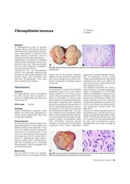
Fig. 1.146 A Fibroadenoma showing lobulated, bulging cut surface. B Fibroadenoma with intracanalicular<br />
growth pattern.<br />
pattern and slit like spaces. Variations<br />
depend on the amount of hyalinization<br />
and myxoid change in the stromal component.<br />
Calcification of sclerotic lesions<br />
is common.<br />
Histopathology<br />
The admixture of stromal and epithelial<br />
proliferation gives rise to two distinct<br />
g rowth patterns of no clinical significance.<br />
The pericanalicular pattern is the<br />
result of proliferation of stromal cells<br />
around ducts in a circumferential fashion;<br />
this pattern is observed most frequently<br />
during the second and third decades of<br />
life. The intracanalicular pattern is due to<br />
compression of the ducts into clefts by<br />
the proliferating stromal cells. The stromal<br />
component may sometimes exhibit<br />
focal or diffuse hypercellularity (especially<br />
in women less than 20 years of age),<br />
atypical bizarre multinucleated giant<br />
cells {233,2278}, extensive myxoid<br />
changes or hyalinization with dystrophic<br />
calcification and, rare l y, ossification<br />
A<br />
B<br />
(especially in postmenopausal women).<br />
Foci of lipomatous, smooth muscle<br />
{1040}, and osteochondroid {1852,2762}<br />
metaplasia may rarely occur. Mitotic figures<br />
are uncommon. Total infarction has<br />
been reported, but rarely.<br />
The epithelial component can show a<br />
wide spectrum of typical hyperplasia,<br />
mainly in adolescents {411,1525,1861,<br />
2250}, and metaplastic changes such as<br />
apocrine or squamous metaplasia may<br />
be seen. Foci of fibrocystic change, sclerosing<br />
adenosis and even extensive<br />
myoepithelial proliferation can also occur<br />
in FA. In situ lobular, and ductal <strong>carcinoma</strong><br />
occasionally develop within FA s<br />
{693,1525}.<br />
Juvenile (or cellular) fibroadenomas are<br />
characterized by increased stromal cellularity<br />
and epithelial hyperplasia {1861,<br />
2250}. The term giant FA has been used<br />
as a synonym for juvenile fibroadenoma<br />
by some, but is restricted to massive<br />
fibroadenomas with usual histology by<br />
others.<br />
Fig. 1.147 Juvenile fibroadenoma. A Lobulated sectioned surface in a 8 cm tumour. Patient was 16 years old.<br />
B Periductal growth pattern with moderate stromal hypercellularity.<br />
B<br />
Fibroepithelial tumours<br />
99


