

Invasive breast carcinoma - IARC
Invasive breast carcinoma - IARC
Invasive breast carcinoma - IARC

You also want an ePaper? Increase the reach of your titles
YUMPU automatically turns print PDFs into web optimized ePapers that Google loves.
1 tumours are extremely rare. Almost<br />
invariably high grade (grade 3) nuclei<br />
are identified. In view of this finding as<br />
well as the mixture of different architectural<br />
patterns, clear cell adeno<strong>carcinoma</strong><br />
is not graded.<br />
Prognosis and predictive factors<br />
When controlled for stage, survival of<br />
women with clear cell adeno<strong>carcinoma</strong><br />
may be slightly lower than that of patients<br />
with serous <strong>carcinoma</strong>. The five year survival<br />
is 69% for patients with stage I<br />
tumours, 55% for stage II, 14% for stage<br />
III and 4% for stage IV. There is no consensus<br />
in the literature about the value of<br />
pattern, cell type, mitotic index or grade<br />
as a prognostic indicator {395}.<br />
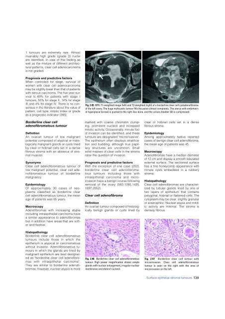
B<br />
Fig. 2.45 MRI: T1-weighted image (left) and T2-weighted (right) of a borderline clear cell cystadenofibroma<br />
of the left ovary. The huge multicystic tumour fills the pelvis almost completely. The uterus with endometrial<br />
hyperplasia (arrow) is pushed to the right iliac bone, and the urinary bladder (B) is compressed.<br />
Borderline clear cell<br />
adenofibromatous tumour<br />
Definition<br />
An ovarian tumour of low malignant<br />
potential composed of atypical or histologically<br />
malignant glands or cysts lined<br />
by clear or hobnail cells set in a dense<br />
f i b rous stroma with an absence of stromal<br />
invasion.<br />
S y n o n y m s<br />
Clear cell adenofibromatous tumour of<br />
low malignant potential, clear cell aden<br />
o f i b romatous tumour of bord e r l i n e<br />
m a l i g n a n c y.<br />
E p i d e m i o l o g y<br />
Of approximately 30 cases of neoplasms<br />
classified as borderline clear<br />
cell adenofibromatous tumour, the mean<br />
age of patients was 65 years.<br />
M a c r o s c o p y<br />
A d e n o f i b romas with increasing atypia<br />
including intraepithelial <strong>carcinoma</strong> have<br />
a similar appearance to adenofibro m a s<br />
but in addition have areas that are softer<br />
and fleshier.<br />
H i s t o p a t h o l o g y<br />
B o rderline clear cell adenofibro m a t o u s<br />
tumours include those in which the<br />
epithelium is atypical or carc i n o m a t o u s<br />
without invasion. Adenofibromatous tumours<br />
in which the glands are lined by<br />
malignant epithelium are best designated<br />
as "borderline clear cell adenofibromas<br />
with intraepithelial carc i n o m a " .<br />
They are similar to borderline adenofib<br />
romas; however, nuclear atypia is more<br />
marked with coarse chromatin clumping,<br />
prominent nucleoli and incre a s e d<br />
mitotic activity. Occasionally, minute foci<br />
of invasion can be identified, and these<br />
tumours are designated "micro i n v a s i v e " .<br />
The epithelium often displays stratification<br />
and budding, although true papill<br />
a ry structures are uncommon. Small<br />
solid masses of clear cells in the stro m a<br />
raise the question of invasion.<br />
Prognosis and predictive factors<br />
With the exception of one case {202},<br />
b o rderline clear cell adenofibro m a -<br />
tous tumours including those with<br />
intraepithelial <strong>carcinoma</strong> and microinvasion<br />
have a benign course following<br />
removal of the ovary {583,1285,1435,<br />
1 8 9 7 , 2 0 5 2 } .<br />
Clear cell adenofibroma<br />
Definition<br />
An ovarian tumour composed of histologically<br />
benign glands or cysts lined by<br />
Fig. 2.46 Borderline clear cell adenofibromatous<br />
tumour. High power magnification shows simple<br />
glands with nuclear enlargement, irregular nuclear<br />
membranes and distinct nucleoli.<br />
clear or hobnail cells set in a dense<br />
fibrous stroma.<br />
Epidemiology<br />
Among approximately twelve reported<br />
cases of benign clear cell adenofibroma,<br />
the mean age of patients was 45.<br />
Macroscopy<br />
Adenofibromas have a median diameter<br />
of 12 cm and display a smooth lobulated<br />
external surface. The sectioned surface<br />
has a fine honeycomb appearance with<br />
minute cysts embedded in a rubbery<br />
stroma.<br />
Histopathology<br />
Clear cell adenofibromas are characterized<br />
by tubular glands lined by one or<br />
two layers of epithelium that contains<br />
polygonal, hobnail or flattened cells. The<br />
cytoplasm may be clear, slightly granular<br />
or eosinophilic. Nuclear atypia and mitotic<br />
activity are minimal. The stroma is<br />
densely fibrous.<br />
Fig. 2.47 Borderline clear cell tumour with<br />
microinvasion. Clear cell adenofibromatous<br />
tumour is seen on the right with the area of<br />
microinvasion on the left.<br />
Surface epithelial-stromal tumours 139


