- Page 2:
Previous volumes in this series Kle
- Page 6 and 7:
World Health Organization Classific
- Page 8 and 9:
Published by IARC Press, Internatio
- Page 10 and 11:
CHAPTER 1 Tumours of the Breast Can
- Page 12 and 13:
TNM classification of carcinomas of
- Page 14 and 15:
Invasive breast carcinoma I.O. Elli
- Page 16 and 17:
Rapid growth and greater adult heig
- Page 18 and 19:
tives, administrative and clerical
- Page 20 and 21:
Grade 1 - well differentiated: 3-5
- Page 22 and 23:
ent and this is occasionally extens
- Page 24 and 25:
Most melanotic tumours of the breas
- Page 26 and 27:
A Fig. 1.22 A Classic invasive lobu
- Page 28 and 29:
yet others at 90% {97,2147}. For pr
- Page 30 and 31:
Fig. 1.27 Medullary carcinoma. The
- Page 32 and 33:
myoepithelial cells in fibro a d e
- Page 34 and 35:
A Fig. 1.33 Neuroendocrine carcinom
- Page 36 and 37:
Macroscopy Fisher et al. reported t
- Page 38 and 39:
Fig. 1.39 Apocrine carcinoma. Note
- Page 40 and 41:
cells contain intracytoplasmic lume
- Page 42 and 43:
A Fig. 1.50 Carcinosarcoma. A Two a
- Page 44 and 45:
A B C Fig. 1.75 Lobular neoplasia.
- Page 46 and 47:
Intraductal proliferative lesions F
- Page 48 and 49:
A B absence of either microcalcific
- Page 50 and 51:
Fig. 1.83 Atypical ductal hyperplas
- Page 52 and 53:
A B Fig. 1.88 Large excision biopsy
- Page 54 and 55:
unusual variants as well. Using thi
- Page 56 and 57:
esemble those identified in invasiv
- Page 58 and 59:
A B Fig. 1.97 Microinvasive carcino
- Page 60 and 61:
A Papilloma may be subject to morph
- Page 62 and 63:
A B C Fig. 1.103 Papillary intraduc
- Page 64 and 65:
colour measuring from 0.5 to 12 cm.
- Page 66 and 67:
Immunoprofile The cases studied by
- Page 68 and 69:
Glycogen-rich, clear cell carcinoma
- Page 70 and 71:
Differential diagnosis There may be
- Page 72 and 73:
quality metaphase spreads from an i
- Page 74 and 75:
Fig. 1.67 Lobular carcinoma of brea
- Page 76 and 77:
detectable distant metastasis displ
- Page 78 and 79:
detected at a late stage as small t
- Page 80 and 81:
Extent of ductal carcinoma in situ
- Page 82 and 83:
Benign epithelial proliferations G.
- Page 84 and 85:
Fig. 1.112 Microglandular adenosis.
- Page 86 and 87:
A Apocrine adenoma ICD-O code 8401/
- Page 88 and 89:
A B Fig. 1.128 Adenomyoepithelioma,
- Page 90 and 91:
Mesenchymal tumours M. Drijkoningen
- Page 92 and 93:
1113,1275}. There is a complex patt
- Page 94 and 95:
A B Fig. 1.137 A Lipoma. Intraparen
- Page 96 and 97:
Fig. 1.141 Angiosarcoma after breas
- Page 98 and 99:
A Fig. 1.144 Mammary osteosarcoma.
- Page 100 and 101: Fibroepithelial tumours J.P. Belloc
- Page 102 and 103: aged women (average age of presenta
- Page 104 and 105: lished data suggests a 21% rate of
- Page 106 and 107: A Fig. 1.157 Syringomatous adenoma
- Page 108 and 109: Malignant lymphoma and metastatic t
- Page 110 and 111: used. Inflammatory conditions in th
- Page 112 and 113: male population both in the USA and
- Page 114 and 115: CHAPTER 2 Tumours of the Ovary and
- Page 116 and 117: Polyembryoma 9072/3 Non-gestational
- Page 118 and 119: Surface epithelial-stromal tumours
- Page 120 and 121: A Fig. 2.04 A Serous borderline tum
- Page 122 and 123: A Fig. 2.06 Serous borderline tumou
- Page 124 and 125: A Fig. 2.10 Invasive peritoneal imp
- Page 126 and 127: Fig. 2.15 Mucinous carcinoma with i
- Page 128 and 129: Fig. 2.20 Mucinous endocervical-lik
- Page 130 and 131: A Fig. 2.28 Mucinous cystic tumour
- Page 132 and 133: early secretory endometrium {2605}.
- Page 134 and 135: IV, 6% {2233}. Patients with grade
- Page 136 and 137: A Fig. 2.37 A This polypoid intracy
- Page 138 and 139: Fig. 2.40 Endometrioid cyst with at
- Page 140 and 141: 1 tumours are extremely rare. Almos
- Page 142 and 143: Fig. 2.50 Borderline Brenner tumour
- Page 144 and 145: express thrombomodulin and have bee
- Page 146 and 147: serous and transitional cell carcin
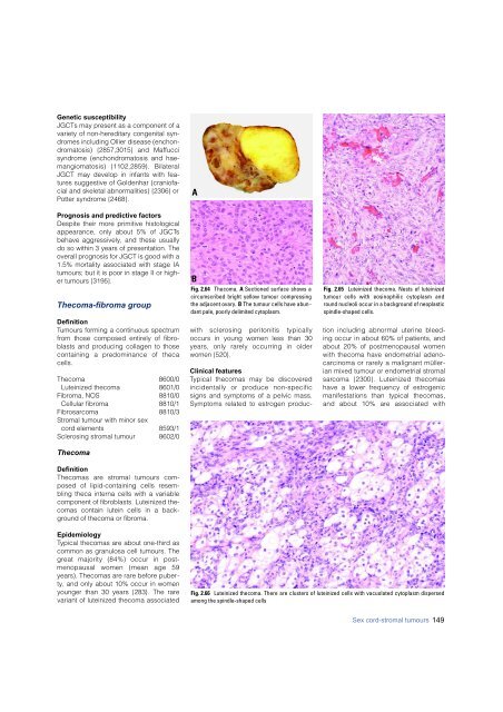
- Page 148 and 149: is yellow to tan with a variable ad
- Page 152 and 153: Clinical features F i b romas may b
- Page 154 and 155: distinguishes this tumour from the
- Page 156 and 157: A Fig. 2.77 A Retiform Sertoli-Leyd
- Page 158 and 159: Mean ages of 21 and 38 years and me
- Page 160 and 161: Tumours unassociated with PJS form
- Page 162 and 163: mal origin without crystals of Rein
- Page 164 and 165: Germ cell tumours F. Nogales A. Tal
- Page 166 and 167: cases, anisokaryosis has no prognos
- Page 168 and 169: ation may coexist in the same neopl
- Page 170 and 171: Table 2.06 Grading of ovarian immat
- Page 172 and 173: A B Fig. 2.102 A Mature cystic tera
- Page 174 and 175: Diagnostic procedures Elevated urin
- Page 176 and 177: associated with mature teratomas sh
- Page 178 and 179: atives intimately admixed. The germ
- Page 180 and 181: Fig. 2.113 Mixed germ cell-sex cord
- Page 182 and 183: A Fig. 2.116 A Adenomatous hyperpla
- Page 184 and 185: Fig. 2.117 Small cell carcinoma, hy
- Page 186 and 187: Clinical features Adenoid cystic-li
- Page 188 and 189: Histopathology This epithelial tumo
- Page 190 and 191: sis. Corpus luteum of pregnancy has
- Page 192 and 193: Lymphomas and leukaemias L.M. Roth
- Page 194 and 195: Secondary tumours of the ovary J. P
- Page 196 and 197: Occasionally, the colonic adenocarc
- Page 198 and 199: Peritoneal tumours S.C. Mok J.O. Sc
- Page 200 and 201:
A B Fig. 2.140 Cystic adenomatoid m
- Page 202 and 203:
whelmingly poor {1038,1547,2310}. H
- Page 204 and 205:
CHAPTER 3 Tumours of the Fallopian
- Page 206 and 207:
TNM and FIGO classification of carc
- Page 208 and 209:
Endometrioid adenocarcinoma Endomet
- Page 210 and 211:
Two examples of adenofibroma of bor
- Page 212 and 213:
A Fig. 3.11 Adenomatoid tumour. A T
- Page 214 and 215:
Clinical features Patients range in
- Page 216 and 217:
Fig. 3.16 Uterus-like mass. The cys
- Page 218 and 219:
CHAPTER 4 Tumours of the Uterine Co
- Page 220 and 221:
TNM and FIGO classification of non-
- Page 222 and 223:
Epithelial tumours and related lesi
- Page 224 and 225:
endometrioid adenocarcinoma fro m a
- Page 226 and 227:
fers from the prototypical type I e
- Page 228 and 229:
the ovary and bladder. Unlike prima
- Page 230 and 231:
schema {1535,2602}. Although this c
- Page 232 and 233:
Table 4.03 Altered gene function in
- Page 234 and 235:
Mesenchymal tumours and related les
- Page 236 and 237:
areas are limited to less than 30%
- Page 238 and 239:
A B C Fig. 4.30 Leiomyosarcoma. A T
- Page 240 and 241:
e used sparingly and is reserved fo
- Page 242 and 243:
A B Fig. 4.38 Leiomyoma with perino
- Page 244 and 245:
cytological atypia, tumour cell nec
- Page 246 and 247:
Mixed epithelial and mesenchymal tu
- Page 248 and 249:
Prognosis and predictive factors Th
- Page 250 and 251:
tumours may superficially invade th
- Page 252 and 253:
A Fig. 4.46 A Gestational choriocar
- Page 254 and 255:
A Fig. 4.49 A Classic complete hyda
- Page 256 and 257:
Sex cord-like, neuroectodermal and
- Page 258 and 259:
Secondary tumours of the uterine co
- Page 260 and 261:
WHO histological classification of
- Page 262 and 263:
Epithelial tumours M. Wells J.M. Ne
- Page 264 and 265:
Fig. 5.04 Mechanisms of human papil
- Page 266 and 267:
Fig. 5.08 Keratinizing squamous cel
- Page 268 and 269:
in their Asian population {2957}. I
- Page 270 and 271:
have a strong association with high
- Page 272 and 273:
e red by squamous epithelium {380}.
- Page 274 and 275:
endometrioid adenocarcinoma of the
- Page 276 and 277:
metrial epithelium. In some cases t
- Page 278 and 279:
with distinct cell borders and a gr
- Page 280 and 281:
Mesenchymal tumours M.L. Carcangiu
- Page 282 and 283:
{781}, liposarcoma {2840,3016}, ost
- Page 284 and 285:
Mixed epithelial and mesenchymal tu
- Page 286 and 287:
Fig. 5.44 Wilms tumour. The tumour
- Page 288 and 289:
A Fig. 5.47 Implant of endometrial
- Page 290 and 291:
CHAPTER 6 Tumours of the Vagina Alt
- Page 292 and 293:
Epithelial tumours E.S. Andersen A.
- Page 294 and 295:
Aetiology The fact that both VAIN a
- Page 296 and 297:
Fig. 6.10 Fibroepithelial polyp.A m
- Page 298 and 299:
er of mitoses varies but is usually
- Page 300 and 301:
ICD-O codes Adenosquamous carcinoma
- Page 302 and 303:
A C Fig. 6.17 Sarcoma botryoides. A
- Page 304 and 305:
1-11 cm. They may arise anywhere in
- Page 306 and 307:
elsewhere in the female genital tra
- Page 308 and 309:
A Fig. 6.24 Yolk sac tumour. A The
- Page 310 and 311:
g rowing in sheets. Some may have s
- Page 312 and 313:
WHO histological classification of
- Page 314 and 315:
Epithelial tumours E.J. Wilkinson M
- Page 316 and 317:
even with additional sectioning, it
- Page 318 and 319:
type) is a highly diff e rentiated
- Page 320 and 321:
Clinical features Bartholin gland c
- Page 322 and 323:
glandular elements surrounded by fi
- Page 324 and 325:
Mesenchymal tumours R.L. Kempson M.
- Page 326 and 327:
Table 7.03 Differential diagnosis o
- Page 328 and 329:
Prognosis and predictive factors A
- Page 330 and 331:
Table 7.04 Clark levels of cutaneou
- Page 332 and 333:
A Fig. 7.23 Vulvar peripheral primi
- Page 334 and 335:
Familial aggregation of cancers of
- Page 336 and 337:
BRCA1 syndrome Definition Inherited
- Page 338 and 339:
dispose individuals to the developm
- Page 340 and 341:
Fig. 8.05 To assess whether wild-ty
- Page 342 and 343:
Fig. 8.07 Factors that modify risk
- Page 344 and 345:
BRCA2 syndrome R. Eeles S. Piver S.
- Page 346 and 347:
tubal or ovarian carcinomas. Howeve
- Page 348 and 349:
in typical nuclear foci that may re
- Page 350 and 351:
Breast tumours Frequency Breast can
- Page 352 and 353:
Fig. 8.14 Codon distribution of som
- Page 354 and 355:
Breast tumours Age distribution and
- Page 356 and 357:
Hereditary non-polyposis colon canc
- Page 358 and 359:
essary in the evaluation of the pat
- Page 360 and 361:
Age distribution and penetrance Mos
- Page 362 and 363:
Contributors Dr Vera M. ABELER** De
- Page 364 and 365:
Dr Carlo LA VECCHIA Laboratory of E
- Page 366 and 367:
Dr Manuel TEIXEIRA Department of Ge
- Page 368 and 369:
02.100 Dr. A. Ostor 02.101 Dr. F.A.
- Page 370 and 371:
52. Akhtar M, Robinson C, Ashraf Al
- Page 372 and 373:
180. Bapat K, Brustein S (1989). Ut
- Page 374 and 375:
307. Bolis GB, Maccio T (2000). Cle
- Page 376 and 377:
436. Chang J, Sharpe JC, A'Hern RP,
- Page 378 and 379:
562. Costa MJ, Ames PF, Walls J, Ro
- Page 380 and 381:
689. Di Domenico A, Stangl F, Benni
- Page 382 and 383:
820. Falck J, Petrini JH, Williams
- Page 384 and 385:
943. Gad A, Azzopardi JG (1975). Lo
- Page 386 and 387:
1067. Grimes MM (1992). Cystosarcom
- Page 388 and 389:
1197. Herod JJ, Shafi MI, Rollason
- Page 390 and 391:
1323. Jacques SM, Qureshi F, Ramire
- Page 392 and 393:
1449. Khalifa MA, Mannel RS, Harawa
- Page 394 and 395:
1564. Lagios MD (1977). Multicentri
- Page 396 and 397:
1687. Loman N, Johannsson O, Kristo
- Page 398 and 399:
1807. McCluggage G, McBride H, Maxw
- Page 400 and 401:
1937. Mukai M, Torikata C, Iri H (1
- Page 402 and 403:
2056. Norris HJ, Taylor HB (1967).
- Page 404 and 405:
2180. Park JS, Jones RW, McLean MR,
- Page 406 and 407:
2304. Puls LE, Hamous J, Morrow MS,
- Page 408 and 409:
2 4 3 4 . Rosen PP, Groshen S, Saig
- Page 410 and 411:
2559. Schlesinger C, Silverberg SG
- Page 412 and 413:
2686. Silverberg SG (1999). Protoco
- Page 414 and 415:
2806. Stutz JA, Evans AJ, Pinder S,
- Page 416 and 417:
2942. Tornos C, Silva EG, Ordonez N
- Page 418 and 419:
3061. Wargotz ES, Norris HJ (1990).
- Page 420 and 421:
3189. Yoshioka T, Tanaka T (2000).
- Page 422 and 423:
References 425
- Page 424 and 425:
Biphasic teratomas, 168 BLM, 341, 3
- Page 426 and 427:
G GADD45, 343, 353 GCDFP-15, 25, 33
- Page 428 and 429:
MPNST, see Malignant peripheral ner
- Page 430:
Small cell / oat cell carcinoma, 32