

Invasive breast carcinoma - IARC
Invasive breast carcinoma - IARC
Invasive breast carcinoma - IARC

You also want an ePaper? Increase the reach of your titles
YUMPU automatically turns print PDFs into web optimized ePapers that Google loves.
DIN terminology is used, the traditional<br />
t e rminology should be mentioned as<br />
well. The classification of intraductal proliferative<br />
lesions should be viewed as an<br />
evolving concept that may be modified<br />
as additional molecular genetic data<br />
become available.<br />
Diagnostic reproducibility<br />
Multiple studies have assessed re p roducibility<br />
in diagnosing the range of intraductal<br />
proliferative lesions, some with<br />
emphasis on the borderline lesions {299,<br />
5 0 3 , 2 1 5 5 , 2 1 5 7 , 2 4 1 1 , 2 5 7 1 , 2 7 2 3 , 2 7 2 4 } .<br />
These studies have clearly indicated that<br />
i n t e robserver agreement is poor when no<br />
s t a n d a rdized criteria are used {2411}.<br />
Although diagnostic re p roducibility is<br />
i m p roved with the use of standardized criteria<br />
{2571} discrepancies in diagnosis<br />
persist in some cases, particularly in the<br />
distinction between ADH and limited form s<br />
of low grade DCIS. In one study, consistency<br />
in diagnosis and classification did<br />
not change significantly when interpre t a-<br />
tion was confined to specific images as<br />
c o m p a red with assessment of the entire<br />
tissue section on a slide, reflecting inconsistencies<br />
secondary to diff e rences in<br />
morphological interpretation {780}. While<br />
clinical follow-up studies have generally<br />
demonstrated increasing levels of bre a s t<br />
cancer risk associated with UDH, ADH<br />
and DCIS re s p e c t i v e l y, concerns about<br />
diagnostic re p roducibility have led some<br />
to question the practice of utilizing these<br />
risk estimates at the individual level {299}.<br />
Aetiology<br />
In general, the factors that are associated<br />
with the development of invasive bre a s t<br />
c a rcinoma are also associated with<br />
i n c reased risk for the development<br />
of intraductal proliferative lesions {1439a,<br />
1551a,2536a}. (See section on epidemiology<br />
of <strong>breast</strong> carc i n o m a ) .<br />
Genetics of precursor lesions<br />
To date, several genetic analyses have<br />
been perf o rmed on potential pre c u r s o r<br />
lesions of <strong>carcinoma</strong> of the <strong>breast</strong>. The<br />
sometimes contradictory results (see<br />
below) may be due to: (i) small number of<br />
cases analysed, (ii) the use of diff e re n t<br />
histological classification criteria, (iii) histomorphological<br />
heterogeneity of both the<br />
n o rmal and neoplastic <strong>breast</strong> tissue and<br />
(iv) genetic hetero g e n e i t y, as identified by<br />
either conventional cytogenetics {1175}<br />
or by fluorescence in situ hybridization<br />
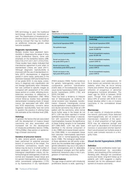
Table 1.11<br />
Classification of intraductal proliferative lesions.<br />
Traditional terminology<br />
Usual ductal hyperplasia (UDH)<br />
Flat epithelial atypia<br />
Atypical ductal hyperplasia (ADH)<br />
Ductal <strong>carcinoma</strong> in situ,<br />
low grade (DCIS grade 1)<br />
Ductal <strong>carcinoma</strong> in situ,<br />
intermediate grade (DCIS grade 2)<br />
Ductal <strong>carcinoma</strong> in situ,<br />
high grade (DCIS grade 3)<br />
(FISH) analysis {1949}. Further evidence<br />
for genetic heterogeneity comes from<br />
comparative genomic hybridization<br />
(CGH) data of microdissected tissue in<br />
usual ductal hyperplasia (UDH), atypical<br />
ductal hyperplasia (ADH) {135} and<br />
DCIS {134,366}.<br />
T h e re has been a tendency to interpre t<br />
loss of heterozygosity as evidence for<br />
clonal evolution and neoplastic transformation.<br />
However, histologically norm a l<br />
ductal epithelium closely adjacent to invasive<br />
ductal <strong>carcinoma</strong> may share an LOH<br />
p a t t e rn with the <strong>carcinoma</strong>, while norm a l<br />
ducts further away in the <strong>breast</strong> do not<br />
{671}. LOH has been re p o rted in norm a l<br />
epithelial tissues of the <strong>breast</strong>, in association<br />
with <strong>carcinoma</strong> and in re d u c t i o n<br />
mammoplasties, however, the significance<br />
of these finding remains to be evaluated<br />
{671,1586,1945}. LOH has also been<br />
identified in the stromal component of<br />
in situ {1889} and invasive <strong>breast</strong> carc i n o-<br />
ma {1545,1889}, in non-neoplastic tissue<br />
f rom reduction mammoplasty specimens<br />
{1568}, and in normal-appearing bre a s t<br />
ducts {1586}. The biological significance<br />
of these alterations are still poorly understood,<br />
but the available data suggest that<br />
genetic alterations may occur very early in<br />
b reast tumorigenesis prior to detectable<br />
morphological changes and that epithel<br />
i a l / s t romal interactions play a role in prog<br />
ression of mammary carc i n o m a .<br />
Clinical features<br />
The age range of women with intraductal<br />
proliferative lesions is wide, spanning 7<br />
Ductal intraepithelial neoplasia (DIN)<br />
terminology<br />
Usual ductal hyperplasia (UDH)<br />
Ductal intraepithelial neoplasia,<br />
grade 1A (DIN 1A)<br />
Ductal intraepithelial neoplasia,<br />
grade 1B (DIN 1B)<br />
Ductal intraepithelial neoplasia,<br />
grade1C (DIN 1C)<br />
Ductal intraepithelial neoplasia,<br />
grade 2 (DIN 2)<br />
Ductal intraepithelial neoplasia,<br />
grade 3 (DIN 3)<br />
to 8 decades post adolescence. All<br />
these lesions are extremely rare prior to<br />
puberty; when they do occur among<br />
infants and children, they are generally a<br />
reflection of exogenous or abnorm a l<br />
endogenous hormonal stimulation. The<br />
mean age for DCIS is between 50-59<br />
years. Though most often unilateral,<br />
about 22% of women with DCIS in one<br />
<strong>breast</strong> develop either in situ or invasive<br />
c a rcinoma in the contralateral bre a s t<br />
{3055}.<br />
Macroscopy<br />
A vast majority of intraductal pro l i f e r a-<br />
tive lesions, particularly those detected<br />
m a m m o g r a p h i c a l l y, are not evident on<br />
m a c roscopic inspection of the specimen.<br />
A small pro p o rtion of high grade<br />
DCIS may be extensive enough and<br />
with such an abundance of intraluminal<br />
n e c rosis or associated stromal re a c t i o n<br />
that it would present as multiple areas of<br />
round, pale comedo necrosis or a firm ,<br />
gritty mass.<br />
Usual ductal hyperplasia (UDH)<br />
Definition<br />
A benign ductal proliferative lesion typically<br />
characterized by secondary<br />
lumens, and streaming of the central proliferating<br />
cells. Although not considered<br />
a precursor lesion, long-term follow-up of<br />
patients with UDH suggests a slightly<br />
elevated risk for the subsequent development<br />
of invasive <strong>carcinoma</strong>.<br />
64 Tumours of the <strong>breast</strong>


