

Invasive breast carcinoma - IARC
Invasive breast carcinoma - IARC
Invasive breast carcinoma - IARC

Create successful ePaper yourself
Turn your PDF publications into a flip-book with our unique Google optimized e-Paper software.
Malignant lymphoma<br />
and metastatic tumours<br />
J. Lamovec<br />
A. Wotherspoon<br />
J. Jacquemier<br />
Malignant lymphoma<br />
D e f i n i t i o n<br />
Malignant lymphoma of the <strong>breast</strong> may<br />
p resent as a primary or secondary<br />
tumour; both are rare. There is no morphological<br />
criterion to diff e re n t i a t e<br />
between the two {117,1792}.<br />
The criteria for defining and documentation<br />
of primary <strong>breast</strong> lymphoma, first<br />
p roposed by Wiseman and Liao {3136}<br />
and, with minor modifications, accepted<br />
by others, are as follows:<br />
1. Availability of adequate histological<br />
material.<br />
2. Presence of <strong>breast</strong> tissue in, or adjacent<br />
to, the lymphoma infiltrate.<br />
3. No concurrent nodal disease except<br />
for the involvement of ipsilateral axillary<br />
lymph nodes.<br />
4. No previous history of lymphoma<br />
involving other organs or tissues.<br />
As such criteria seem too restrictive and<br />
leave no room for primary <strong>breast</strong> lymphomas<br />
of higher stages, some authors<br />
include cases in which the <strong>breast</strong> is<br />
the first or major site of pre s e n t a t i o n ,<br />
even if, on subsequent staging proc<br />
e d u res, involvement of distant nodal<br />
sites or bone marrow is discovere d<br />
{ 3 5 9 , 1 2 6 1 , 1 7 5 3 } .<br />
Epidemiology<br />
P r i m a ry <strong>breast</strong> lymphoma may appear at<br />
any age, but the majority of patients are<br />
postmenopausal women. A subset of<br />
patients is re p resented by pregnant or lactating<br />
women with massive bilateral bre a s t<br />
swelling; most of these cases were re p o rted<br />
from Africa {2643} although non-<br />
African cases are also on re c o rd {1753}.<br />
The disease is exceedingly rare in men<br />
{ 2 5 4 0 } .<br />
Clinical features<br />
Clinical presentation of primary <strong>breast</strong><br />
lymphoma usually does not differ from<br />
that of <strong>breast</strong> <strong>carcinoma</strong>. It usually presents<br />
with a painless lump sometimes<br />
multinodular, which is bilateral in approximately<br />
10% of cases. Imaging usually<br />
reveals no feature which helps to distinguish<br />
primary from secondary lymphoma<br />
{1657,2199}. The value of MR imaging in<br />
<strong>breast</strong> lymphomas has not been clearly<br />
determined {1952,1961}.<br />
Macroscopy<br />
P r i m a ry and secondary <strong>breast</strong> lymphomas<br />
most commonly appear as a<br />
well circumscribed tumour of vary i n g<br />
size, up to 20 cm in largest diameter. On<br />
cut surface, the neoplastic tissue is white<br />
to white-grey, soft or firm, with occasional<br />
haemorrhagic or necrotic foci {994,<br />
1580,1753,3136}.<br />
Histopathology<br />
Microscopically, the majority of primary<br />
<strong>breast</strong> lymphomas are diffuse large B<br />
cell lymphomas, according to the most<br />
recent WHO classification {352,1144}. In<br />
older literature, cases designated as<br />
reticulum cell sarcoma, histiocytic lymphoma<br />
and at least some lymphosarcoma<br />
cases would nowadays most probably<br />
be included in the above category.<br />
M o re re c e n t l y, such lymphomas were<br />
diagnosed as centroblastic or immunoblastic<br />
by the Kiel classification or diffuse<br />
large cell cleaved or noncleaved<br />
and immunoblastic lymphomas by the<br />
Lukes-Collins classification and Working<br />
F o rmulation {18,296,534,706,994,1261,<br />
1346,1580,1665}.<br />
A minor pro p o rtion of primary lymphomas<br />
of the <strong>breast</strong> reflect Burkitt lymphoma,<br />
extranodular marginal-zone B-<br />
cell lymphoma of mucosa associated<br />
lymphoid tissue (MALT) type, follicular<br />
lymphoma, lymphoblastic lymphoma of<br />
either B or T type, and, extremely rarely,<br />
T-cell lymphomas of variable subtypes<br />
by the current WHO classification.<br />
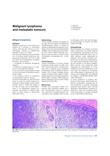
A<br />
Fig. 1.159 Diffuse large B-cell lymphoma. A Medullary <strong>carcinoma</strong>-like appearance. B Circumscribed mass, composed of large pleomorphic neoplastic lymphoid<br />
cells.<br />
B<br />
Malignant lymphoma and metastatic tumours 107


